Fig. 3.

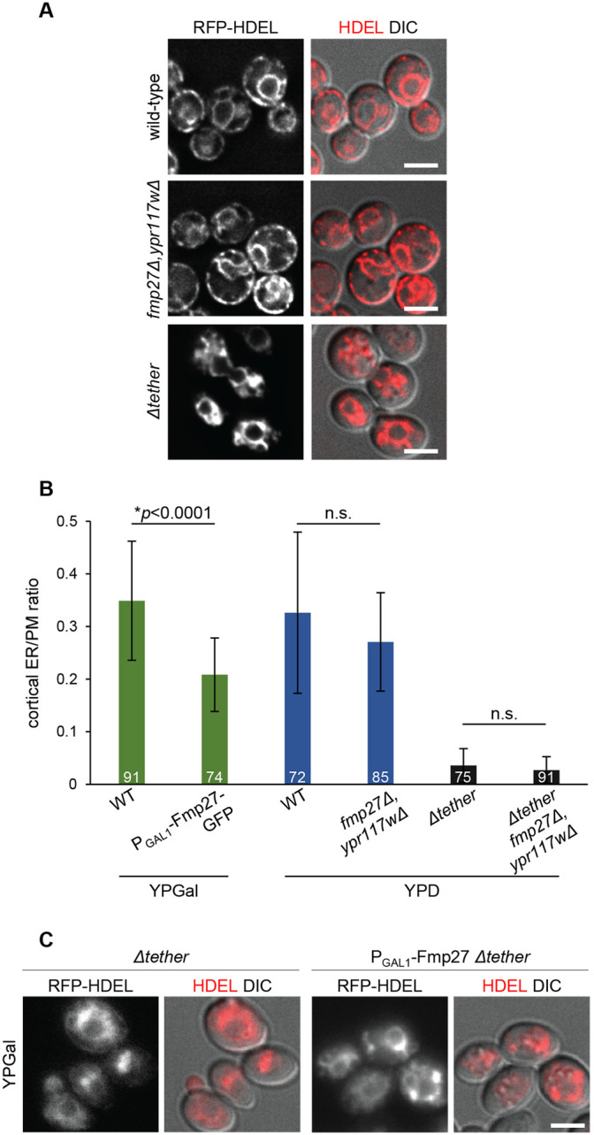
Fmp27 does not function as an ER-PM tether. (A) Live-cell imaging of RFP–HDEL (red) in control, fmp27Δ ypr117wΔ and Δtether cells showing that ER morphology is unaffected upon loss of both copies of the yeast ortholog of hobbit, in contrast to Δtether cells, where nearly all cortical ER is lost. (B) Quantification of the ratio of cortical ER to total PM in control (wild-type, WT), FMP27–GFP overexpression, fmp27Δ ypr117wΔ, Δtether, and Δtether fmp27Δ ypr117wΔ cells showing that overexpression of Fmp27 does not increase the number of cortical ER contacts (green bars); in fact, the number of contacts is significantly reduced. Loss of both Fmp27 and Ypr117w does not decrease the number of cortical ER contacts in a wild-type (blue bars) or Δtether background (gray bars). Cells were grown on YPGal medium to drive Fmp27 overexpression; otherwise, cells were grown on standard YPD medium. Graphs show mean±s.d.; statistics calculated by Welch's t-test; n.s., not significant. The number on each bar represents the number of cells for which the cortical-ER to PM ratio was measured. (C) Live-cell imaging of RFP–HDEL (red) in Δtether (left) and Δtether cells overexpressing Fmp27 (right) showing that overexpression of Fmp27 does not rescue cortical ER contacts in the Δtether background. Fmp27 is expressed under its endogenous promoter in the left panels and under the GAL1 promoter in right panels. Cells were collected from mid-log cultures grown in YPGal medium to drive Fmp27 overexpression. Images shown in A and C are representative of three independent experiments with n≥3 images acquired per replicate. Scale bars: 2 µm.
