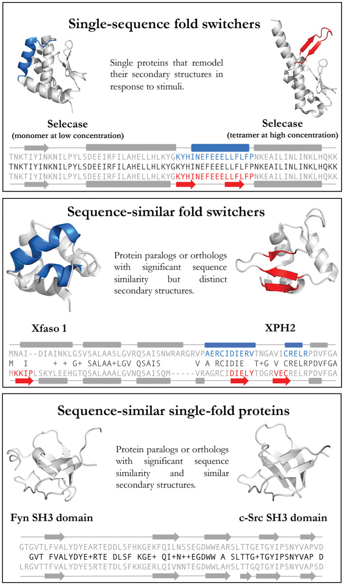
FIGURE 1.

Definitions of single‐sequence fold switchers, sequence‐similar fold switchers, and sequence‐similar single‐fold proteins. Gray regions are structurally unchanged between the two conformations. Upper/lower sequences and secondary structures (rounded rectangle = helix, arrow = strand, line = coil) correspond to protein structures on the left/right, respectively. Middle sequences (black) show amino acid identities (letters) and similarities (+). PDB IDs (Left to right, top to bottom): 4QHF, 4QHH, 3BD1, 5W8Z, 1FYN, 6XVM. Cartoon diagrams were made in PyMOL[ 7 ]
