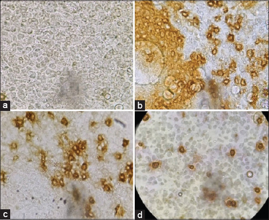
Figure-1.

Thymus and bursa of Fabricius sections showing (a) isotype control. (b) Bursa of Fabricius cell showing lymphocytes expressing Bu-1 marker (brown in color). (c) Thymocytes expressing CD4 marker (brown-stained cells). (d) Thymocytes expressing CD8 marker (brown-stained cells). Primary antibody and biotinylated horse anti-mouse IgG secondary antibody were the first to treat the section. A ”charged” DAB (Abcam, USA) was the primary stain to develop color and methyl green was the counterstain.
