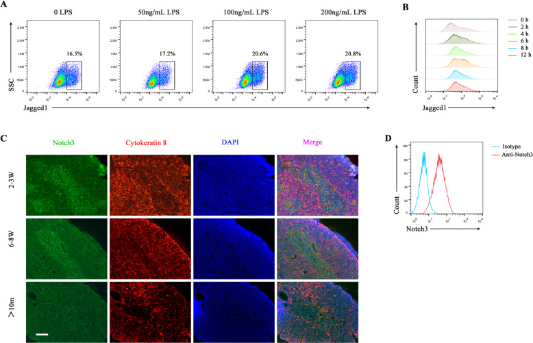
Fig. 3. Mature BMDCs expressed a high level of Jagged1, whereas mTECs expressed Notch3.
A BMDCs were stained with the anti-Jagged1 antibody. The expression level of Jagged1 in BMDCs was examined using FCM after stimulation with different concentrations of LPS for 6 h. B The expression level of Jagged1 in BMDCs after stimulation with LPS (100 ng/ml) for different durations was detected using FCM. C Representative images of the expression of Notch3 (green fluorescence) and cytokeratin 8 (K8, a cTEC marker, red fluorescence) in the thymus from different aged mice (2–3 weeks old, 6–8 weeks old, and 10 months old, respectively) were captured by immunofluorescence microcopy. Paraffin sections of the thymus obtained from different aged mice were stained with primary rabbit anti-Notch3 and rat anti-cytokeratin 8 antibodies, followed by secondary Alexa Fluor 488-labeled goat anti-rabbit and Alexa Fluor 555-labeled goat anti-rat antibodies, respectively. Finally, the sections were stained with DAPI. Scale bars represent 200 µm. D mTEC1 cells were stained with PE-conjugated anti-Notch3 antibody. The expression of Notch3 in mTEC1 cells was detected by FCM. All data are from three independent experiments. Representative figures of these three experiments are displayed.

