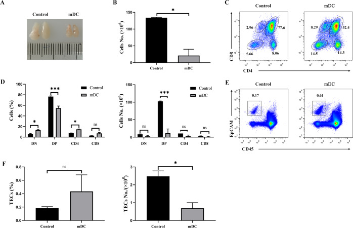
Fig. 5. Intrathymic injection of mature DCs led to acute thymic atrophy.
For A to F: Thymic atrophy following the intrathymic injection of mDCs (n = 3). The same volume of PBS was used as a control (n = 3). A Thymus morphology and size. B Total numbers of thymocytes were calculated. C Thymocytes were stained with PE-conjugated anti-CD4 and APC-labeled anti-CD8 antibodies. The thymocyte subpopulations were examined by FCM. D The percentages and absolute numbers of indicated thymocyte subpopulations were calculated. E The cells isolated from the thymus of recipient mice were stained with Alexa Fluor 488-conjugated anti-CD45 and APC-labeled anti-EpCAM antibodies, respectively. CD45-EpCAM+ TECs were detected by FCM. F The percentages and absolute numbers of CD45-EpCAM+ TECs were calculated. All data are from three independent experiments. Representative figures of these three experiments are shown. Data are expressed as the mean ± SD. *P < 0.05, ***P < 0.001 compared with the control group.

