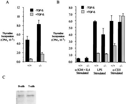
FIG. 5.
Assay of TGF-β’s effects in primary splenocytes reveals both Smad3-dependent and Smad3-independent growth-inhibitory signaling pathways. (A) Smad3 is not required for TGF-β-mediated growth inhibition in primary unstimulated splenocytes. Primary splenocytes were isolated from 8-week-old mice and cultured in the presence or absence of 100 pM TGF-β for 48 h. Cells were incubated with [3H]thymidine for the last 4 h of culture, after which the splenocytes were harvested and 3H incorporation was measured. Bars indicate the average of three identically treated wells for each growth condition; error bars represent the standard deviation. (B) Smad3 is required for TGF-β-mediated growth inhibition of αCD3-stimulated splenocytes. Primary splenocytes were isolated from 8-week-old mice and cultured in the presence of the indicated growth stimuli in the presence or absence of 100 pM TGF-β. Cellular proliferation was assayed by [3H]thymidine incorporation as for panel A. (C) Smad3 is expressed in both B and T cells. Western blotting for Smad3 was performed on purified B and T cells from mature wild-type spleens.

