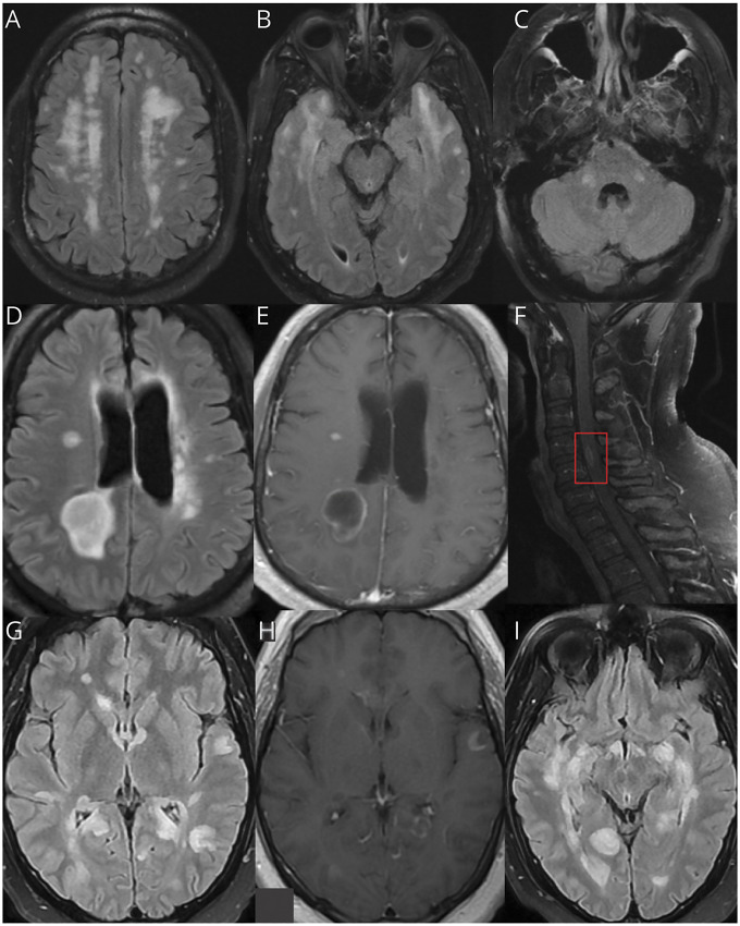
Figure 2. Neuroimaging Examples From the MGB Case Series.
Axial brain MRI sequences of patients with post-COVID ADEM from the MGB case series. (A–C) Axial brain MRI sequences from case 1 display FLAIR hyperintensities in the bilateral frontal lobes, anterior temporal lobes, and cerebellum. (D and E) Axial brain MRI sequences from case 2 show bilateral FLAIR hyperintensities with a prominent right parietal lesion that enhances, as well as nodular enhancement of the right corona radiata and occipital horn of the right lateral ventricle. (F) Sagittal postcontrast T1 sequence of the cervical spine MRI from case 2 displays a focus of intramedullary enhancement at C4-5 (red box). (G–I) Axial brain MRI sequences from case 3 show diffuse, multifocal T2 FLAIR lesions (G and I) and a predominant open-ring contrast enhancement pattern of several lesions in the T1 postcontrast sequence (H). ADEM = acute disseminated encephalomyelitis; MGB = Massachusetts General Brigham.

