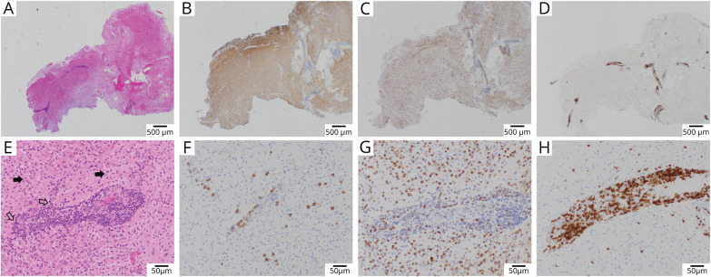
Figure 3. Neuropathology Examples of Post–COVID-19 ADEM.
Brain pathology from case 2 of the MGB series (case details provided in the supplementary files, links.lww.com/NXI/A584). Low-power images (A–D) demonstrate markedly cellular brain parenchyma with prominent perivascular inflammation and reduced myelinated fibers (A, Luxol–H&E stain), with relative preservation of axons (B, neurofilament immunohistochemistry). The cellular population is composed largely of macrophages (C, CD68 immunohistochemistry), whereas the perivascular infiltrates are predominantly T cells (D, CD3 immunohistochemistry). Higher-power views (E–H) characterize the perivascular infiltrates as composed of small mature lymphocytes and focally prominent plasma cells (E, H&E, open arrows), whereas the surrounding tissue is composed of numerous foamy macrophages (solid arrows) and reactive astrocytes. Immunohistochemistry demonstrates scattered perivascular and parenchymal plasma cells (F, CD138), numerous parenchymal foamy macrophages (G, CD68), and marked perivascular T cells (H, CD3). ADEM = acute disseminated encephalomyelitis; MGB = Massachusetts General Brigham.

