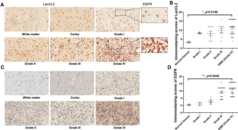
Fig. 5.
Protein expression and intracellular localization of LanCL2 was correlated with the grade of gliomas. A Immunohistochemistry analysis of LanCL2 in representative sections of grade I to IV gliomas. Sections of white matter and cortex were used for comparison. Bar = 20 μm. B IHC staining scores of LanCL2 in tumor sections. C Immunohistochemistry analysis of EGFR in representative sections of grade I to IV gliomas. Bar = 20 μm. D IHC staining scores of EGFR in tumor sections. P values were determined by Kruskal–Wallis One-Way ANOVA and Dunn’s multiple comparisons. *p < 0.05; **p < 0.01

