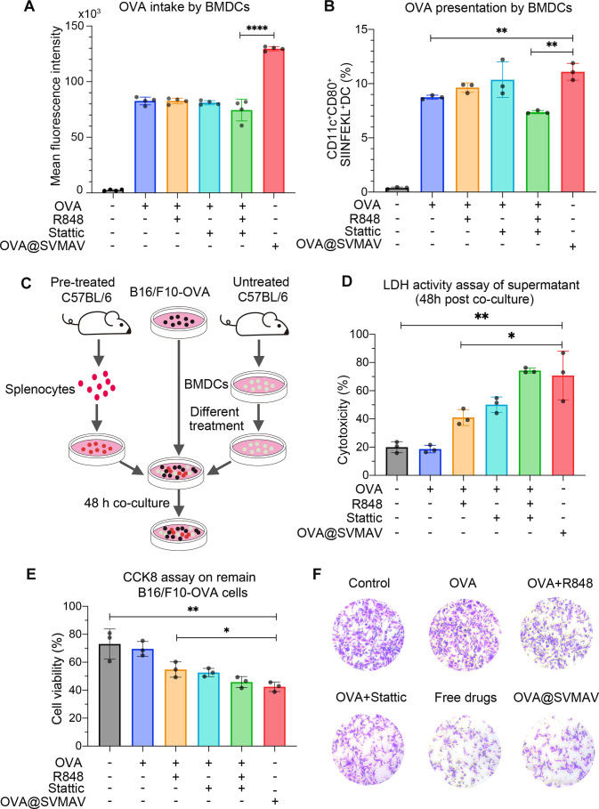
Figure 3.
SVMAV promoted DC functions in vitro. (A) FITC fluorescence intensity of BMDCs with different treatments. (B) Frequency of CD11c+CD80+SIINFEKL+ BMDCs after different treatments. (C) Schematic illustration of the experimental design for cytolytic activity analysis of splenocytes primed by BMDCs. (D) The cytotoxicity of BMDCs treated with different formulations of vaccines was assessed by the LDH assays. (E) The viability of the remaining B16/F10-OVA cells was measured by the CCK8 assay. (F) Representative Photographs of the remaining B16/F10-OVA cells stained with crystal violet for different groups. Data are displayed as mean±SD. *P<0.05, **p<0.01, ****p<0.0001. BMDCs, bone marrow-derived dendritic cells; DC, dendritic cell; FITC, fluorescein isothiocyanate; LDH, lactate dehydrogenase; OVA, ovalbumin; SVMAV, self-assembling vehicle-free multicomponent antitumor nanovaccine.

