Figure 3.
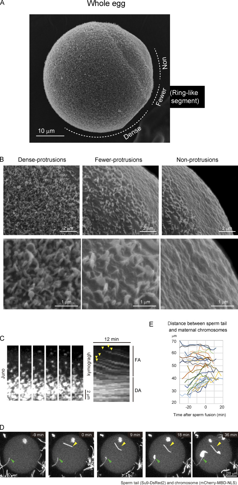
The densities of protrusions on the egg surface correlate with Juno/CD9 structures. (A) Protrusions on the egg surface visualized by scanning electron microscopy (n = 21). The zona pellucida was removed in A–C by treatment with collagenase. (B) High-magnification images in dense-, fewer-, and nonprotrusion areas. In the dense-protrusion area, there are at least two types of structures: one is a finger-like shape, and the other is a round shape. In the fewer-protrusion area, the shape of protrusions is flat. (C) Live imaging of Juno/CD9 structures with fluorescent-labeled Juno and CD9 primary antibodies (n = 15, data of CD9 are not depicted). The Juno/CD9 structures in the FA region moved directionally toward the FA/DA border. The structures in the DA region are relatively stable. A time-lapse video is shown in Video 3. (D) Live imaging of chromosomes and sperm tails. Arrowheads indicate the initial binding position of sperm tails. Two holes were made in the zona pellucida near the actin cap on purpose in D and E. (E) The distance between the sperm tail and maternal chromosomes. Sperm that bind close to maternal chromosomes move away from maternal chromosomes before sperm fusion.

