Figure 5.
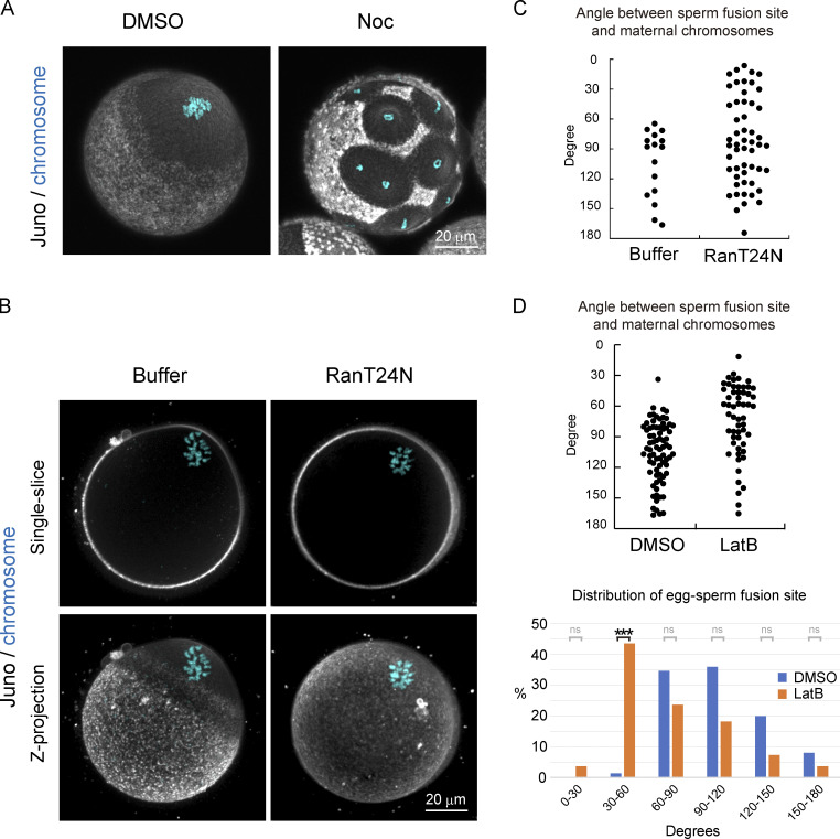
RanGTP activity surrounding maternal chromosomes prevents the localization of Juno and CD9. (A) Immunofluorescence images with fluorescent-labeled Juno and CD9 primary antibodies following treatment with DMSO or Noc (data of CD9 are not depicted). In Noc-treated eggs, Juno/CD9 structures are excluded around individual chromosomes. The zona pellucida was removed in A. (B) Immunofluorescence images with fluorescent-labeled Juno and CD9 primary antibodies in buffer and in RanT24N-injected eggs (data of CD9 is not depicted). In RanT24N-injected eggs, Juno/CD9 structures are formed all over the cell surface, even at regions proximal to maternal chromosomes. (C) Angle between maternal chromosomes and sperm fusion sites in buffer- or RanT24N-injected eggs. In RanT24N-injected eggs, sperm fuse at positions all over the egg surface. Data include zygotes that were polyspermic (polyspermy is 40% [buffer] and 48% [RanT24N] of the fertilized eggs). The zona pellucida was softened by treatment with glutathione. (D) Angle between maternal chromosomes and sperm fusion sites following treatment with DMSO or LatB. LatB-treated eggs exhibited significantly less spatial bias in sperm fusion compared with control eggs. However, sperm still did not fuse around maternal chromosomes. Data include zygotes that were polyspermic (polyspermy is 36% [DMSO] and 44% [LatB] of the fertilized eggs). A hole was made in the zona pellucida using a piezo-driven pipette without glutathione treatment. Fisher’s exact tests were used to obtain P values, and Holm correction was used for the correction of multiple comparison (P value of 30–60° is <8.22 × 10⁻¹⁰; P values of other areas are >0.05 [ns]). ***, P < 0.001.

