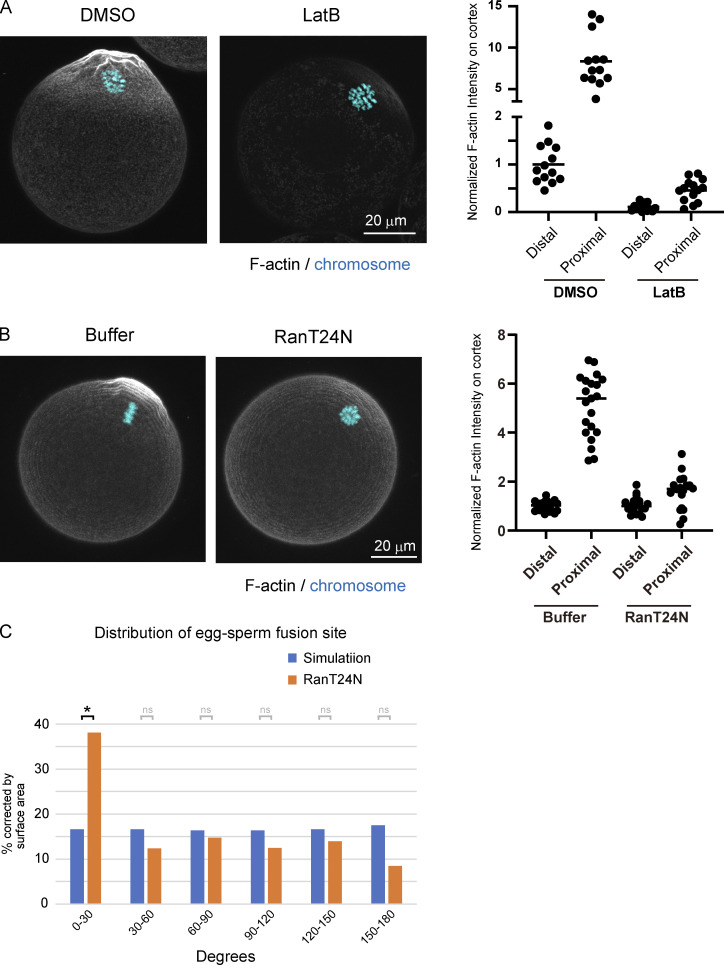
Figure S5.
Actin structures in unfertilized eggs after treatment with LatB. (A) Fluorescent staining of F-actin and chromosomes following treatment with DMSO or LatB. Eggs were fixed and stained with phalloidin and Hoechst 33342. The actin intensity was measured at the cortex near (proximal) or far (distal) from maternal chromosomes in eggs treated with DMSO or LatB. (B) Fluorescent staining of F-actin and chromosomes in Ran-inhibited eggs (RanT24N). The actin intensity was measured at the cortex near (proximal) or far (distal) from maternal chromosomes in Ran-inhibited eggs (RanT24N). (C) Angle between maternal chromosomes and sperm fusion sites in RanT24N-injected eggs. We compare the experimental data with the result of a simulation where sperm fuse randomly anywhere on the egg surface. Data include zygotes that exhibited polyspermy (polyspermy rate was 48% [RanT24N] of the fertilized eggs). Fisher’s exact tests were used to obtain P values, and Holm correction was used for the correction of multiple comparisons (P value of 0–30° is < 3.7 × 10⁻²; P values of other areas are >0.05 [ns]). *, P < 0.05.

