Figure S5.
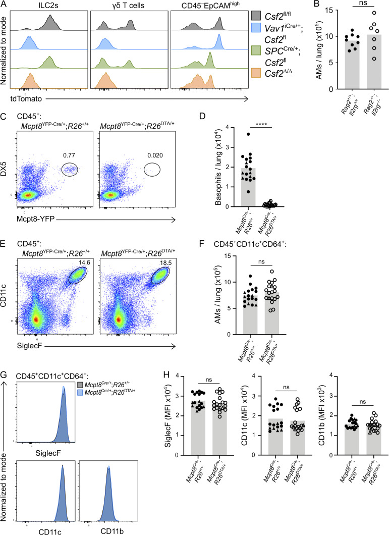
Lung hematopoietic GM-CSF contributions, including from lymphocytes and basophils, are dispensable for AM survival in the adult lung. (A) tdTomato signal from Thy1.2+ST2+ ILC2s, CD3+TCRγδ+ γδ T cells, and CD45−EpCAMhigh cells in adult lungs of Csf2fl/fl (gray), Vav1iCre/+;Csf2fl (blue), SPCCre/+;Csf2fl (green), and Csf2∆/∆ (orange) mice. (B) AM quantification from adult lungs of Rag2+/+;Il2rg+/+ and Rag2−/−;Il2rg−/− mice. (C–H) Analysis of adult lungs isolated from Mcpt8YFP-Cre;R26+/+ and Mcpt8YFP-Cre;R26DTA/+ mice. (D, F, and H) Mcpt8YFP-Cre/+ mice are indicated by circles, while Mcpt8YFP-Cre/YFP-Cre mice are indicated by triangles. (C) Flow cytometry analysis of DX5+Mcpt8-YFP+ basophils, gated on CD45+ cells. (D) Quantification of basophils. (E) Flow cytometry analysis of CD45+CD11c+SiglecF+ AMs. (F) Quantification of CD45+CD11c+CD64+ AMs. (G) Expression levels of SiglecF, CD11c, and CD11b by CD45+CD11c+CD64+ AMs. Mcpt8YFP-Cre/+;R26+/+ (gray) and Mcpt8YFP-Cre/+;R26DTA/+ (blue) mice. (H) MFI of SiglecF, CD11c, and CD11b for CD45+CD11c+CD64+ AMs. (A, C, E, and G) Data are from one experiment representative of four (A) or three (C, E, and G) independent experiments. (B, D, F, and H) Data are pooled from two (B) or three (D, F, and H) independent experiments. ns, P ≥ 0.05; ****, P < 0.0001. MFI, mean fluorescence intensity.

