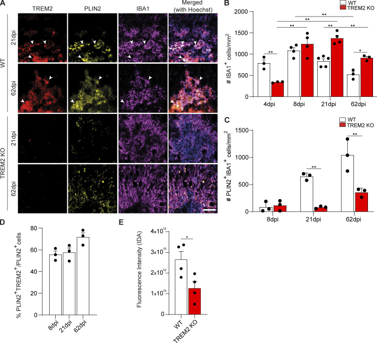
Figure 2.
TREM2 is required for innate immune cell resolution and foam cell formation. (A) Confocal images of 21- and 62-dpi lesions from WT and TREM2 KO mice after staining for TREM2 (red), PLIN2 (yellow), and IBA1 (magenta), showing the colocalization of PLIN2 and TREM2 (arrowheads mark triple-positive cells). Scale bar, 50 μm. (B) Quantification of the density of IBA1+ cells at 4, 8, 21, and 62 dpi. (C) The density of PLIN2+IBA1+ microglia (cells per mm2) was quantified for WT and TREM2 KO. (D) Quantification of the percentage of PLIN2+IBA1+ cells, which are also positive for TREM2, in 21-dpi WT lesions. (E) Quantification of cholesterol crystal fluorescence by reflection confocal microscopy in WT and TREM2 KO lesions at 21 dpi. n = 3–5 animals per condition for A–D; n = 4 lesions per condition for E. Data represent mean ± SEM. For B and C, P values were calculated using two-way ANOVA (A) with Šidák post hoc correction. For D, P values were calculated using one-way ANOVA. For E, P values were calculated using two-tailed unpaired t test. *, P < 0.05; **, P < 0.01. IDA, integrated density area.

