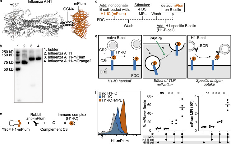
Figure 4.
TLR4 activation on FDCs enhances native antigen presentation. (a) Ribbon representation of H1-mPlum with receptor binding mutant (RBM, Y95F). (b) Western blot of influenza H1 protein expressed with and without fluorescent proteins. (c) Schematic of IC generation. Ig-depleted serum was used as source of complement; for the IC, negative control heat-inactivated serum was used. (d) Timeline of experiment. Sorted cultured FDCs were loaded with influenza H1-IC, washed, and stimulated with MPL or PBS control for 8 h. Medium was replaced, and H1-specific B cells were added for ∼2 h. Then mPlum signal (H1-IC) was measured on H1-specific B cells by flow cytometry. (e) Representation of the experiment. H1-IC on B cells was used as a readout for H1-IC available on the FDC surface. (f) Histogram of mPlum signal on H1-specific B cells with and without MPL stimulation. No mPlum was detected on B cells when FDCs were not loaded with H1-IC or when a nonspecific B cell line was used (NS-B cell, same donor S. pneumoniae–specific). Stimulation with MPL-enhanced specific B cell uptake in percentage as well as MFI. Student’s t test or ANOVA, *, P > 0.05; **, P > 0.01; ***, P value > 0.001; n = 4 (biological replicates). Related to Fig. S4. PAMP, pathogen-associated molecular pattern.

