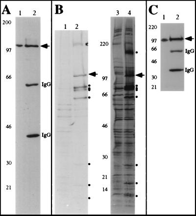
FIG. 3.
lid1p-myc associates with several other proteins in vivo. (A) lid1p-myc is an ∼100-kDa protein. KGY1302 whole-cell lysates (lane 1) or 9E10 immunoprecipitates from KGY1302 lysates (lane 2) were resolved by SDS-PAGE and transferred to a PVDF membrane, and the membrane was probed with the 9E10 antibody. Molecular mass standards (in kilodaltons) are indicated. (B) Wild-type (lanes 1 and 3) or KGY1302 (lid1::lid1-myc) (lanes 2 and 4) native 35S-labeled cell lysates were immunoprecipitated with the 9E10 antibody, and immunoprecipitates were resolved by SDS-PAGE. Labeled proteins from two separate experiments for 1 day (lanes 1 and 2) or 7 days (lanes 3 and 4) were visualized by fluorography. Migrations of molecular mass markers (in kilodaltons) are indicated. Bands specific to the KGY1302 immunoprecipitate are indicated (●). An arrowhead indicates the band corresponding to lid1p-myc. (C) Cut9p-HA is an ∼90-kDa protein. A KGY1365 whole-cell lysate (lane 1) or an HA.11 immunoprecipitate (lane 2) was resolved by SDS-PAGE and transferred to a PVDF membrane, and the membrane was probed with the HA.11 antibody.

