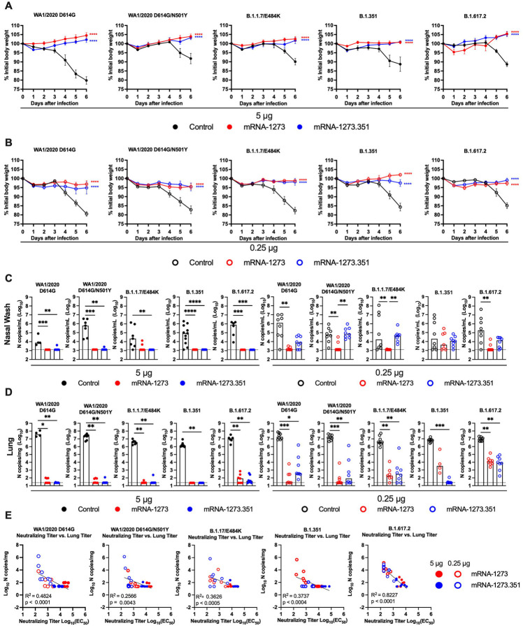
Figure 5. Protection against SARS-CoV-2 infection after mRNA vaccination in K18-hACE2 transgenic mice.
Seven-week-old female K18-hACE2 mice were immunized and boosted with 5 or 0.25 μg of mRNA vaccines (control (black symbols), mRNA-1273 (red symbols), or mRNA-1273.351 (blue symbols) as described in Figure 4A. Four weeks after boosting, mice were challenged via intranasal inoculation with 103 to 3 × 10⁴ FFU of WA1/2020 D614G, WA1/2020 N501Y/D614G, B.1.1.7/E484K, B.1.351, or B.1.617.2, depending on the strain. A-B. Body weight change over time. Data shown is the mean +/− SEM (n = 4–8, two independent experiments; one-way ANOVA of area under the curve from 2–4 dpi with Dunnett’s post-test, comparison to control immunized group: **** P < 0.0001). C-D. Viral burden levels at 6 dpi in the nasal washes (C) and lungs (D) as assessed by qRT-PCR of the N gene after challenge of immunized mice with the indicated mRNA vaccines (n = 4–8, two independent experiments, boxes illustrate median values, dotted line shows LOD; one-way Kruskal-Wallis ANOVA with Dunn’s post-test, comparison among all immunization groups: *, P < 0.05; **, P < 0.01; ***, P < 0.001; **** P < 0.0001). E. Correlation analyses comparing serum neutralizing antibody concentrations three weeks after boosting plotted against lung viral titer (6 dpi) in K18-hACE2 mice after challenge with the indicated SARS-CoV-2 strain (Pearson’s correlation P and R2 values are indicated as insets; closed symbols 5 μg vaccine dose; open symbols, 0.25 μg vaccine dose).

