Figure 6.
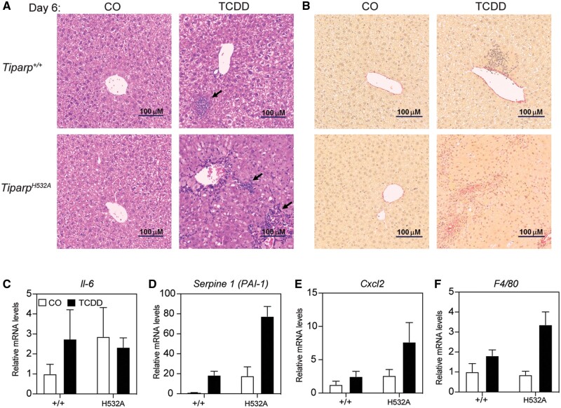
Increased hepatic inflammation in 2,3,7,8-tetrachlorodibenzo-p-dioxin (TCDD)-treated TiparpH532A compared with Tiparp+/+ mice. A, Representative hematoxylin and eosin staining of livers from Tiparp+/+ and TiparpH532A mice (n = 4). The arrows indicate focal inflammatory infiltration. B, Representative picrosirius red staining of livers from Tiparp+/+ and TiparpH532A mice (n = 4). Control animals were injected with CO and were euthanized on day 6. All images are to the same scale. Hepatic gene expression levels of (C) interleukin 6, (D) Serpine 1, (E) Cxcl2, and (F) F4/80 were analyzed as described in the methods. *p < .05 2-way analysis of variance (ANOVA) followed by Sidak’s post hoc test compared with genotyped-matched control-treated mice. #p < .05 2-way ANOVA followed by Sidak’s post hoc test compared with TCDD-treated Tiparp+/+ mice. Figure depicts the mean ± SEM (n = 4).

