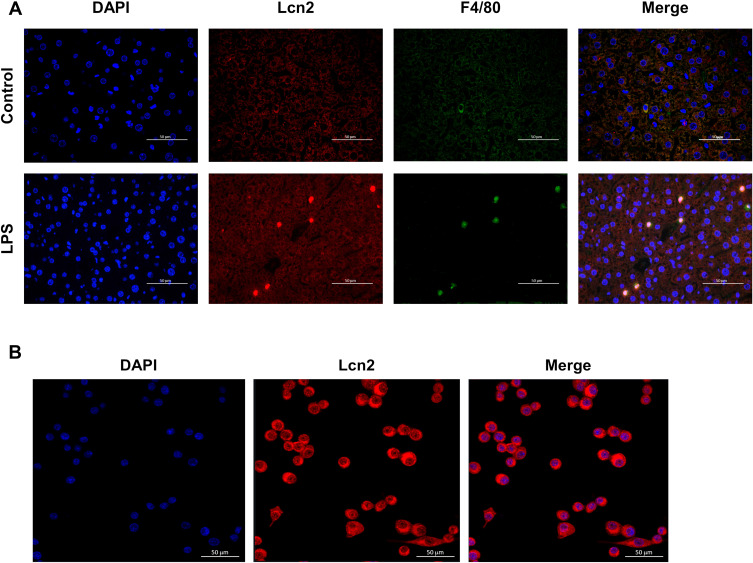
Figure 3.
Lcn2 is predominantly expressed in macrophages during LPS-induced inflammation. (A) Representative confocal images of Lcn2 and macrophages in the liver at 24 h post LPS injection (n = 4). DAPI, blue; Lcn2, red; macrophage marker F4/80, green; Merge, yellow. (B) Representative confocal images of Lcn2 in RAW264.7 macrophages (n = 6). DAPI, blue; Lcn2, red.

