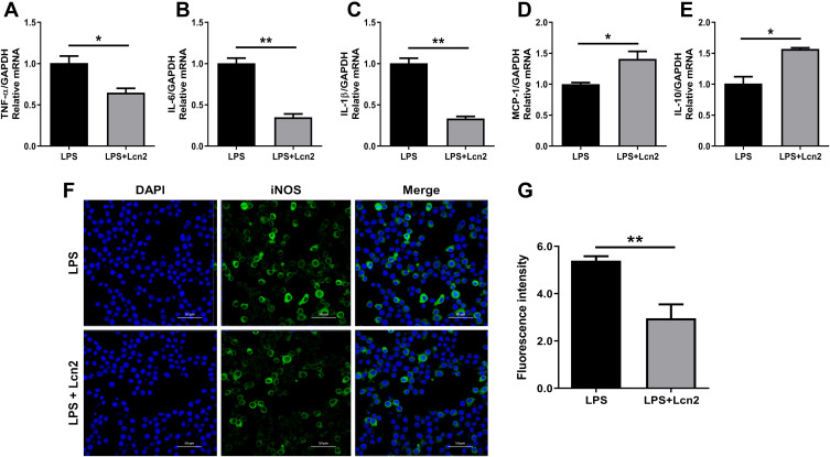
Figure 5.
Lcn2 attenuates the inflammatory response of macrophages. RAW264.7 macrophages were treated with 2 µg/mL recombinant Lcn2 for 22.5 h and 1 µg/mL LPS for 1.5 h. (A–E) Real-time PCR analysis of cytokine mRNA expression levels (n = 3). Relative mRNA expression of cytokines normalized to GAPDH rRNA as the reference genes. The mRNA expression of cytokines in LPS-treated mice was used as the control. (F–G) RAW264.7 macrophages were subjected to staining with rabbit monoclonal antibody iNOS and Alexa Fluor 488 goat anti-rabbit IgG, and observed by laser scanning confocal microscope (n = 3). Cells were assayed for expression by detection of iNOS gene (green) by fluorescence microscopy. Cell nuclei were labeled with DAPI (blue). The number of cells were quantified to the same level for detection of fluorescence intensity. Values were average means of triplicate experiments. Results were expressed as means ± SEM. Statistical analysis used Wilcoxon signed-rank test. *P < 0.05, **P < 0.01.

