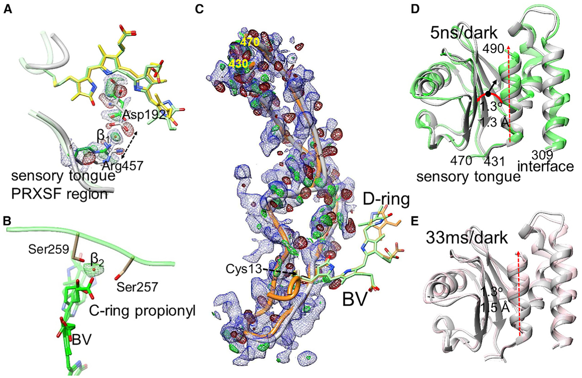
Figure 6. Local and global structural changes.

DED in red and green (±3σ contour), EED in blue (1.2σ contour).
(A) Separation of the sensory tongue from the BV binding region (dotted arrow) in subunit A (5 ns). Asp192 and Arg457 are indicated. The BV chromophore with the 90° twisted and fully isomerized D ring is shown in yellow and green, respectively. The positive DED feature β1 is interpreted by a water molecule.
(B) The BV (green) C-ring propionyl group detaches from Ser257 and Ser259, which coordinate a water (feature β2, green positive DED) instead. The structure is displayed for subunits B (33 ms) where the D ring twists ~90° both clockwise and counterclockwise.
(C) The sensory tongue region. Gray, structure of the reference state; orange, structure at 33 ms. Residues at the beginning and the end of the region are indicated. The chromophore is shown with the twisted D ring (green) and the fully isomerized form (orange).
(D) The PHY domain region. Comparison of the 5-ns structure (green) with the reference structure (gray). Sequence numbers are indicated. The PHY domain centroid (black dot) moves by 1.3 Å (black arrow) and rotates (red curved arrow) by 1.3°. The connection to and from the sensory tongue is marked.
(E) The PHY domain at 33 ms versus reference. Displacements similar to those in (A) are observed. In (D) and (E), the displacement of the C-terminal helix is marked by the red dashed arrow.
