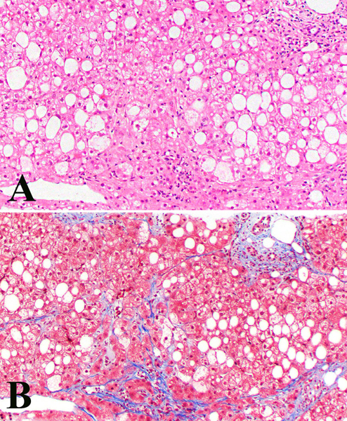
Figure 8.
Medium power view of a liver biopsy demonstrating a steatohepatitic pattern of injury. There is moderate macrovesicular steatosis and hepatocyte ballooning. In this view, there is a focus of lytic necrosis with histiocytes, which may be due to immune checkpoint inhibitor injury, but in this context can look similar to usual non-alcoholic steatohepatitis. Note that the portal tract (upper left) is not inflamed.

