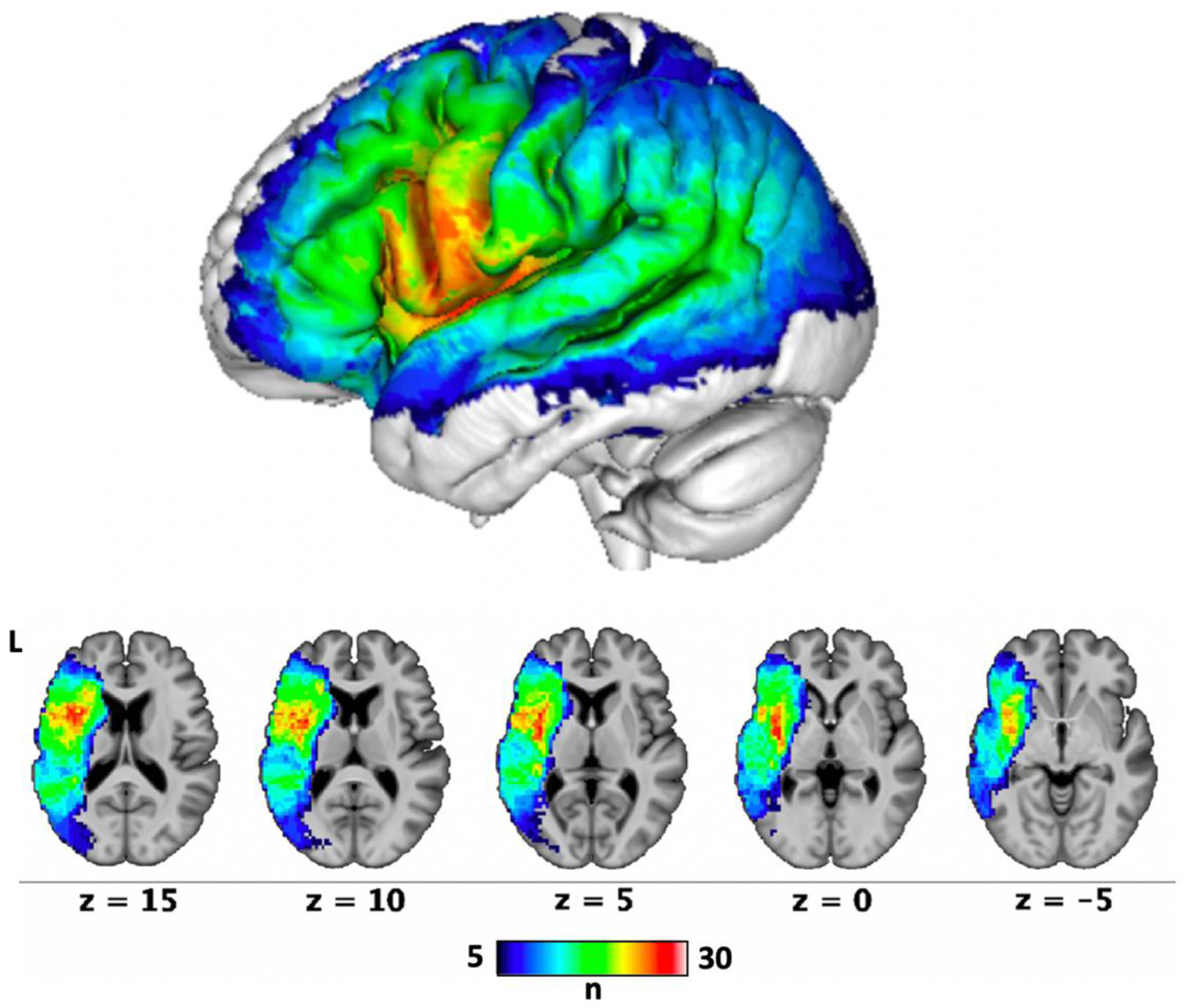
Figure 5. Lesion overlap map (n = 43).

Each voxel is colored based on the number of people with aphasia whose lesions involved that voxel. SVR-LSM analyses were limited to voxels that were lesioned in at least 10% of the participants (n = 5).

Each voxel is colored based on the number of people with aphasia whose lesions involved that voxel. SVR-LSM analyses were limited to voxels that were lesioned in at least 10% of the participants (n = 5).