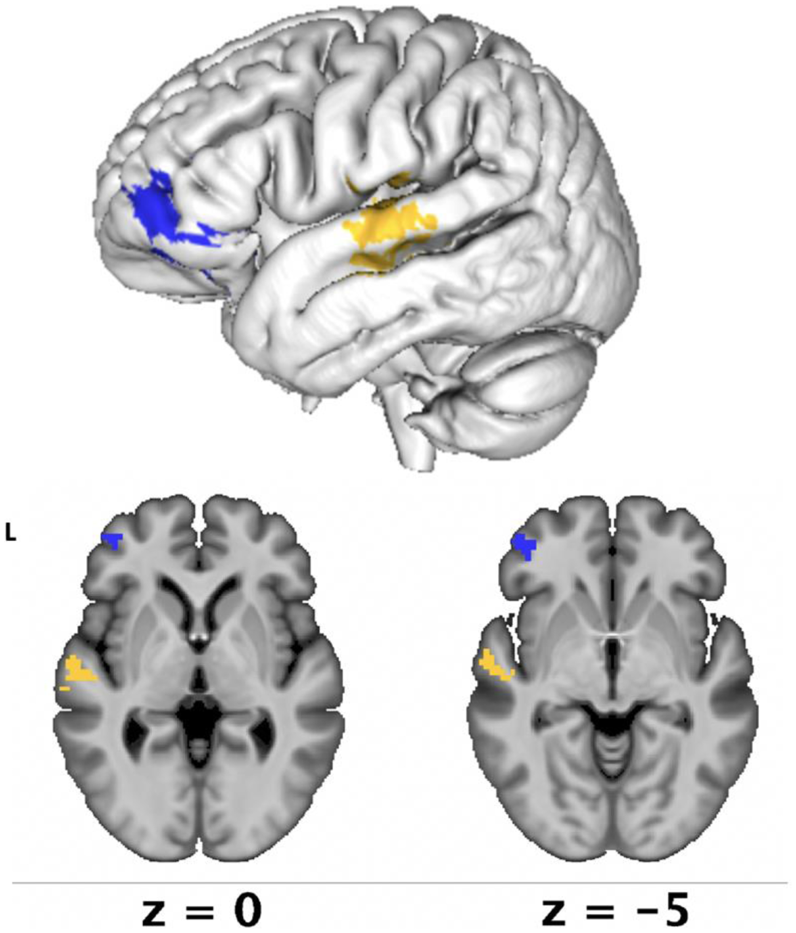
Figure 6. SVR-LSM results for intellectual awareness.

Lesion-symptom mapping results demonstrated that reduced intellectual awareness was associated with lesions in the anterior inferior frontal gyrus, primarily in the pars orbitalis (in blue, p = .03) and preserved intellectual awareness was associated with lesions in the mid-superior temporal gyrus (in yellow, p = .03).
