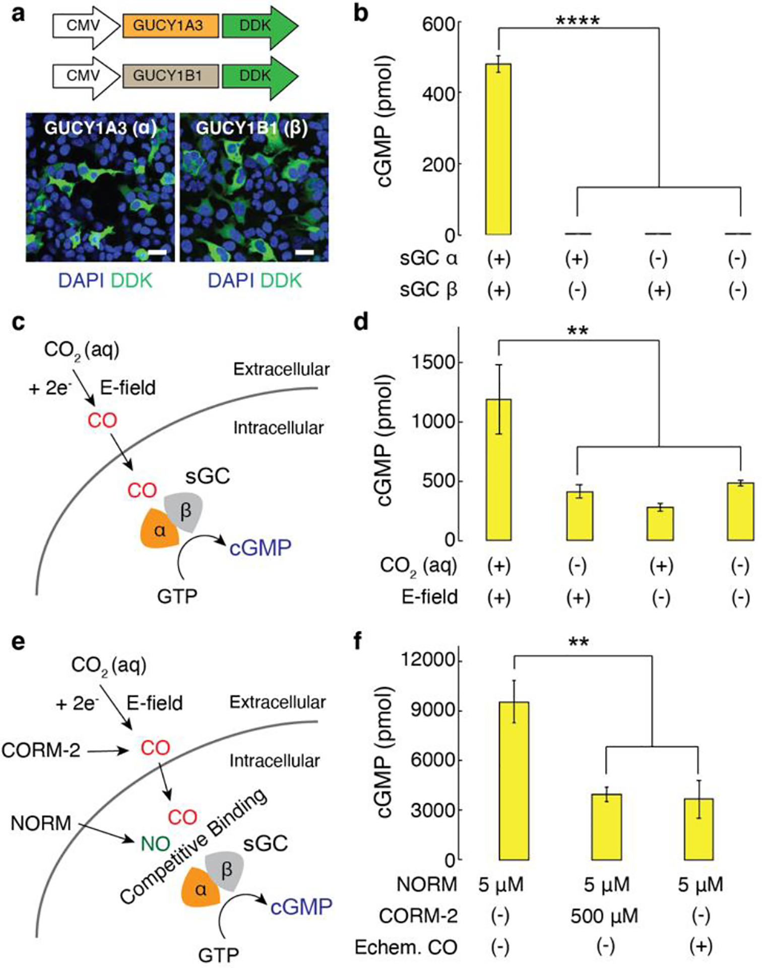
Figure 2.

a, Representative confocal images of HEK cells transfected with DDK-tagged α-subunit of sGC or DDK-tagged β-subunit of sGC under the CMV promoter (scale bar, 50 μm). b, Intracellular cGMP levels (mean ± s.e.m.) in 106 HEK cells 48 h after the transfection (n = 6, one-way analysis of variance (ANOVA) and Tukey’s multiple comparison test, F3,20 = 392.2, **** p = 1.1 × 10⁻¹⁶ < 0.0001). c, A schematic illustrating activation of sGC mediated by electrochemically produced CO. GTP, guanosine 5’ triphosphate. d, Intracellular cGMP levels (mean ± s.e.m.) in 106 sGC+ cells following CO delivery driven by CoPc/OxCP cathodes at −1.3 V versus SHE for 10 min. The statistical significance of an increase in cGMP levels after electrochemical CO delivery as compared with controls was assessed by one-way ANOVA and Tukey’s multiple comparison test (n = 6, F3,20 = 7.4, ** p = 0.0016 < 0.01). e, An illustration of NO-sGC-cGMP signaling pathways modulated by CO. NORM, nitric oxide releasing molecule. f, Intracellular cGMP levels (mean ± s.e.m.) in 106 sGC+ cells following NO delivery in the presence or absence of CO (n = 5, one-way ANOVA and Tukey’s multiple comparison test, F2,12 = 10.9, ** p = 0.002 < 0.01).
