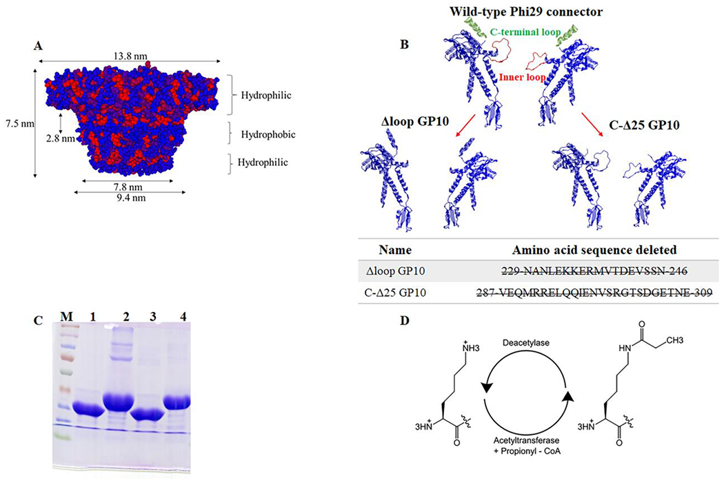
Figure 1. Illustration and characterization of the channel of wild type and mutant phi29 bacterial virus DNA packaging motor.

(A) Side view of the phi29 connector, red hydrophobic; blue hydrophilic. (B) Cross-section structure of two protein subunits of phi29 connector showing the inner loops and the C-terminus and N-terminus. (C) Molecular weight of purified C-Δ25 GP10 (lane 1), N-Δ14 GP10 (lane 2), Δloop GP10 (lane 3) and wild type GP10 (lane 4) on 10% SDS-PAGE; (D) Schematic diagram of lysine propionylation modification.
