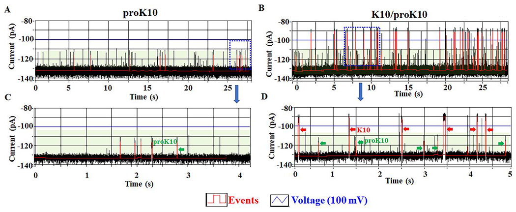
Figure 6. C-Δ25 GP10 channel to distinguish K10 and proK10 on the MinION™ Flow Cell system.

(A, C) The current trace showing the appearance of proRK10 translocation signals (green arrow). (B, D) The current trace showing the appearance of K10 translocation signals (red arrow) after the addition of K10 to MinION™ Flow Cell in the presence of proRK10 (green arrow).
