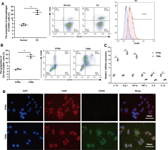
Fig. 1. Accumulation of M2-like TAMs in cancerous tissues of patients with EC.
A Flow cytometry analysis and quantification of macrophages in human EC specimens (n = 3) and normal endometrial specimens (n = 3). Left: CD45+ cells were gated and then analyzed the CD11b+ CD68+ cells. Right: The proportion of CD206+ cells in human EC specimens. B Flow cytometry analysis and quantification of CD11b+ CD206+ macrophages in tissue-resident macrophages (NTMs, n = 3) and tumor-associated macrophages (TAMs, n = 3). C qRT-PCR analysis of IL-10, arginase-1 (Arg-1), transforming growth factor-beta (TGF-β), tumor necrosis factor-alpha (TNF-α), IL-6, and IL-12 mRNA levels in NTMs (n = 3) and TAMs (n = 3). *p < 0.05. D The representative images of immunofluorescent staining for F4/80 (red) and CD206 (green) performed on TAMs and NTMs. Nuclei were counterstained with DAPI (blue). Scale bar = 20 μm.

