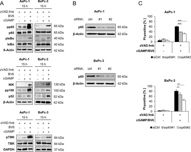
Fig. 6. NF-κB signaling contributes to 2′3′-cGAMP/BV6/zVAD.fmk-induced cell death.
A Western blot analysis of phosphorylated and total p65, IκBα, NIK, p100, p52, and TBK1 in the indicated cell lines treated with 4 µg/ml 2′3′-cGAMP and/or 5 µM BV6 for 15 h in the presence or absence of 20 μM zVAD.fmk. GAPDH or β-Actin serves as loading controls. Representative blots of at least two different independent experiments are shown. B Western blot analysis of p65 in the indicated PC cell lines after 48 h of transfection with non-silencing RNA (ctrl) or siRNA targeting p65. β-Actin serves as loading control. Representative Western blots of at least two different independent experiments are shown. C AsPc-1 and BxPc-3 cells were transfected with control siRNA (siCtrl) or two independent siRNAs targeting p65 and were treated with 4 µg/ml 2′3′-cGAMP and 5 µM BV6 for 48 h in the presence of 20 μM zVAD.fmk. The amount of cell death was calculated by quantifying PI uptake determined with the ImageXpress Micro XLS system. Data are presented as percentage of PI-positive cells, and mean and SD of three independent experiments performed in triplicate are shown. *P < 0.05, **P < 0.01, ***P < 0.001.

