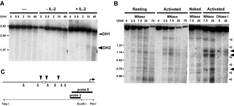
FIG. 7.
IL-2 induces the appearance of DNase I and MNase sites flanking the IL-2rE. (A) DNase I-treated chromatin from ConA-primed cells or primed cells induced further with or without IL-2 for 24 h was digested with TaqI and PstI and electrophoresed in 1% agarose gels. DNase I-hypersensitive (DH) sites were visualized by hybridization with probe 8. DH1 is a lymphocyte-specific constitutive site that maps close to the transcription start site (60). DH2 is located at the IL-2rE. Size markers are indicated on the left (in kilobases). (B) MNase cleavage of naked DNA and chromatin from resting T cells or cells stimulated for 72 h with ConA and IL-2. DNA was digested with TaqI and EcoNI and electrophoresed in a 1.5% agarose gel. The DNase I-treated samples from activated cells in the right panel were digested with the same restriction enzymes. Blots were hybridized with probe 3. Solid arrowheads indicate chromatin-specific cleavage sites in activated cells. Open arrowheads indicate the cutting sites in naked DNA. (C) Summary of MNase analysis. The IL-2rE is represented as a hatched box. Arrowheads (as in panel B) show mean cleavage positions of the MNase cuts deduced from two experiments.

