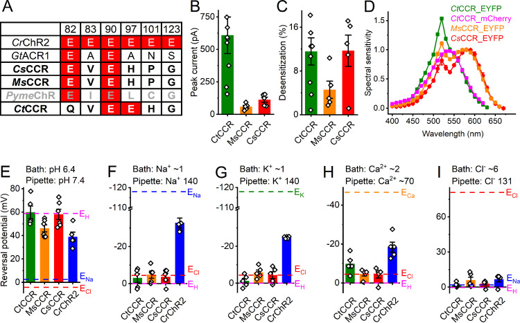
FIG 2.
Prasinophyte CCRs. (A) Amino acid residues corresponding to the indicated positions in CrChR2. ChRs characterized in this study are in bold (black, functional; gray, nonfunctional). Conserved glutamates are highlighted in red. (B) Peak photocurrent amplitudes generated at −60 mV in response to 1-s light pulses at the wavelength of the spectral maximum. (C) Desensitization of photocurrents after 1-s illumination. (D) Action spectra of photocurrents. The data points show means and SEM (n = 4 to 8 scans). (E and I) Reversal potentials of photocurrents. The bars in panels B, C, and E to I show means ± SEM; diamonds show data from individual cells.

