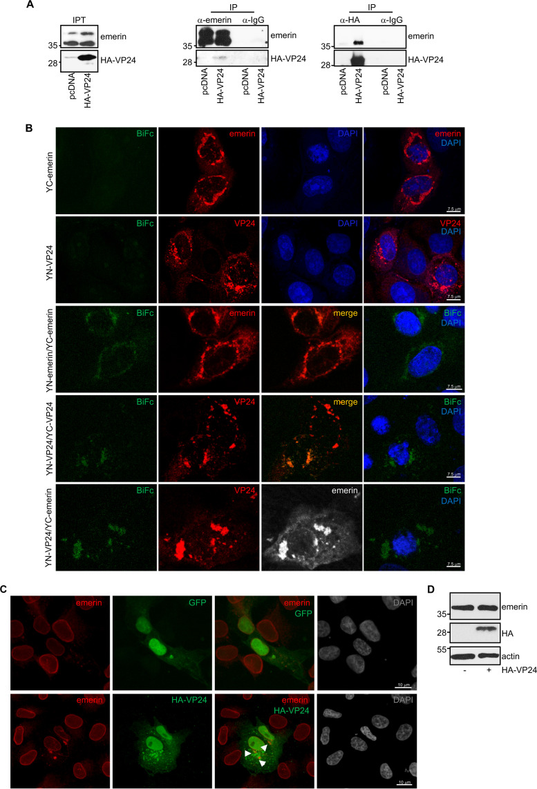
FIG 1.
Interaction of EBOV VP24 protein with emerin. (A) Coimmunoprecipitation between VP24 and emerin. Vero cells seeded in 100-mm plates were transfected with 5 μg of pcDNA or HA-VP24, and 36 h after transfection, protein extracts of transfected cells were immunoprecipitated using anti-emerin, anti-HA, or anti-IgG antibodies. Immunoprecipitated proteins were analyzed by Western blotting using the indicated antibodies. The anti-emerin antibody detected a major band of around 37 kDa and a higher molecular weight band of around 39 kDa, probably corresponding to phosphorylated emerin. The experiments were repeated twice, and representative images of one experiment are shown; IP, immunoprecipitated samples; IPT, input cell extract. (B) VP24-emerin colocalization using the BiFc system. Vero cells were transfected with the indicated combination of the BiFc constructs (YC-emerin, C-terminal part of the yellow fluorescent protein [YFP] fused to the N terminus of full-length emerin; YN-emerin, N-terminal part of YFP fused to the N terminus of full-length emerin; YN-VP24, N-terminal part of YFP fused to the N-terminal part of full-length VP24; YC-VP24, C-terminal part of YFP fused to the N-terminal part of full-length VP24). Cells were fixed, permeabilized, and stained with anti-emerin and/or anti-VP24 primary antibodies. Chromosomes were stained with DAPI (blue). Coexpression of YC- and YN-emerin, YC- and YN-VP24, or YN-VP24 and YC-emerin led to the reconstitution of YFP signal (BiFc). The data represent more than three biological replicates. (C) Localization of endogenous emerin in Vero cells transfected with 0.3 μg of GFP or HA-VP24 or in untransfected cells. Emerin and HA-tagged VP24 are shown. Chromosomes were stained with DAPI. Arrowheads indicate colocalization of HA-VP24 and emerin. (D) Western blotting analysis using anti-emerin antibody of Vero cells at 36 h after transfection with 0.3 μg of pcDNA or HA-VP24 expression plasmids.

