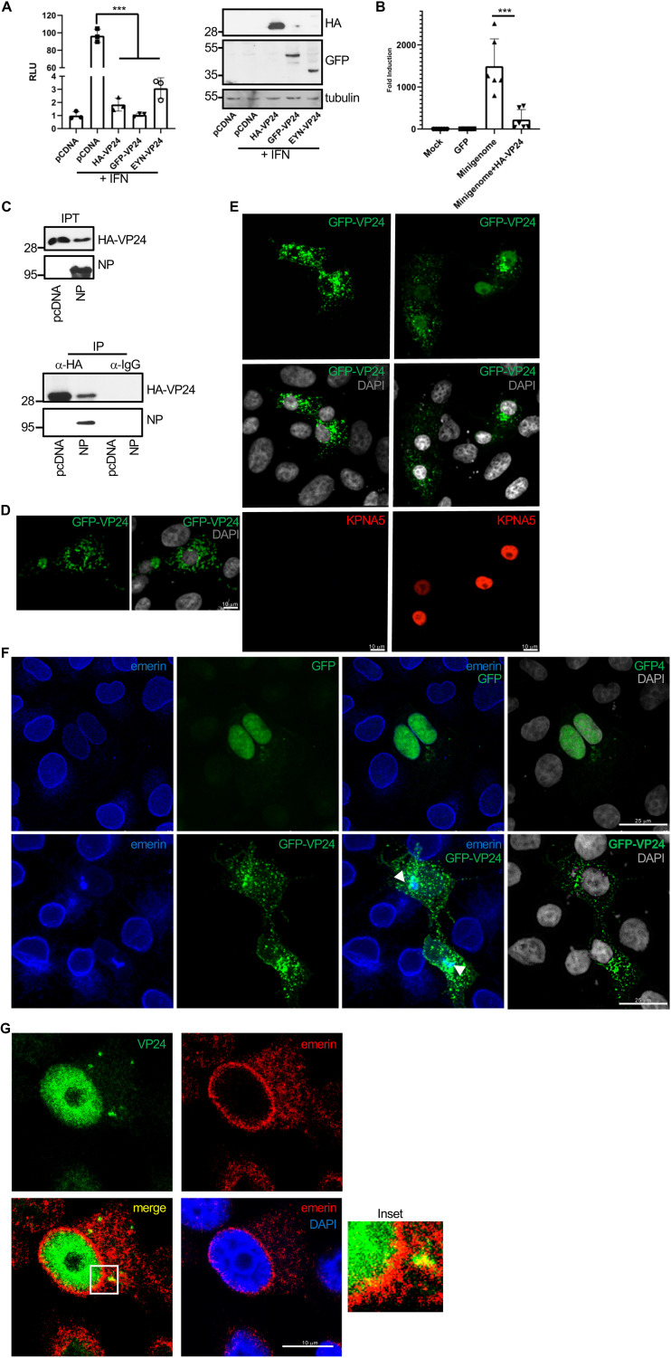
FIG 4.
EBOV VP24 protein interacts with emerin in a tag-independent manner. (A) HEK-293 cells were cotransfected with ISG54-luciferase together with pcDNA or the indicated VP24 expression plasmids. At 24 h after transfection, cells were treated with IFN-α, and at 16 h after treatment, luciferase production was analyzed. Columns are representative of the mean, and error bars represent the standard deviation of three biological replicates (left). Cell lysates from the experiment were analyzed by Western blotting for VP24 expression (right). (B) Minigenome assay in cells cotransfected with HA-VP24. Columns are representative of the mean, and error bars represent the standard deviation of three biological replicates. (C) Coimmunoprecipitation between HA-VP24 and NP. Vero cells were cotransfected with 2.5 μg of HA-VP24 and 2.5 μg of pcDNA or NP expression plasmids in a 100-mm dish, and 36 h after transfection, protein extracts of transfected cells were immunoprecipitated using anti-HA antibody. Immunoprecipitated proteins were analyzed by Western blotting using the indicated antibodies. The experiments were repeated twice, and representative images of one experiment are shown; IP, immunoprecipitated samples; IPT, input cell extract. (D) Localization of GFP-VP24 protein in Vero cells. Chromosomes were stained with DAPI. (E) Localization of GFP-VP24 protein in cells cotransfected with pcDNA or karyopherin 5 (KPNA5) expression plasmid. Chromosomes were stained with DAPI. (F) Colocalization of emerin and GFP-VP24 in Vero cells cotransfected with GFP or GFP-VP24. Chromosomes are stained with DAPI. Arrowheads indicate colocalization of GFP-VP24 and emerin. (G) Colocalization of emerin and VP24 in Vero cells transfected with untagged VP24 expression plasmid. Chromosomes are stained with DAPI. Inset shows higher magnification of the boxed area.

