FIG 7.
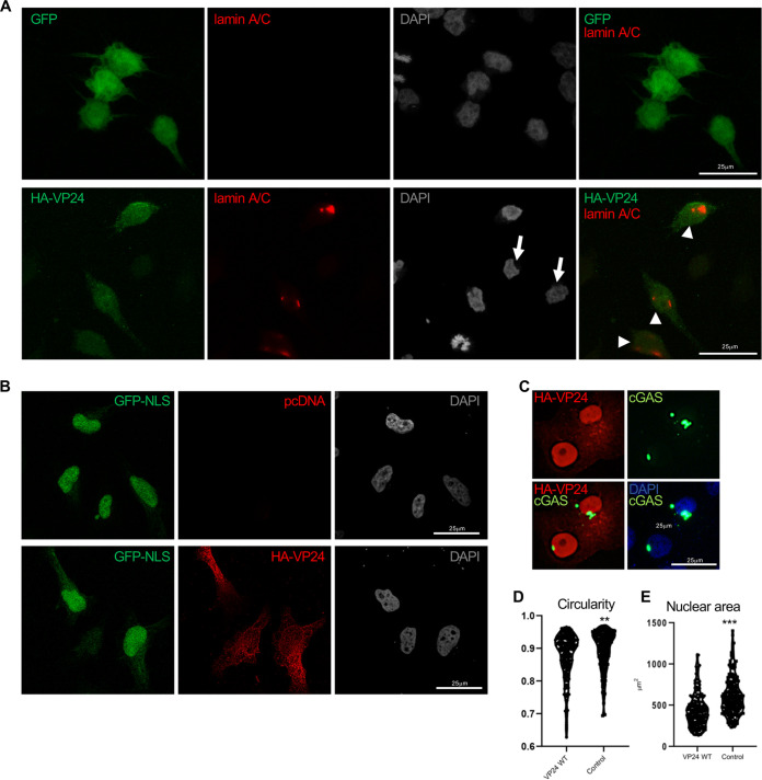
VP24 induces nuclear membrane disruption. (A) Immunofluorescence staining using anti-lamin A/C antibody of cells transfected with 0.3 μg of GFP or VP24 and permeabilized with digitonin. Chromosomes were stained with DAPI. Arrowheads indicate positive detection of lamin A/C in cells expressing VP24, and arrows indicate untransfected cells. (B) Localization of GFP-NLS in cells cotransfected with 0.3 μg of pcDNA or HA-VP24 and permeabilized with digitonin. Chromosomes were stained with DAPI. (C) Localization of GFP-cGAS in Vero cells transfected with 0.3 μg of HA-VP24. (D) Shape of the nucleus of Vero cells expressing HA-VP24 or control cells. A higher circularity denotes a more circular shape. (E) Size of the nucleus of Vero cells expressing HA-VP24 or control cells. Graphs show one data point per nucleus analyzed. Statistical analysis was assessed by a Student’s t test. **, P < 0.01; ***, P < 0.001.

