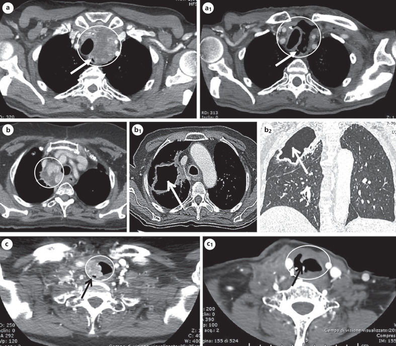
Fig. 2.
CT images of some cases who developed fistula and/or organ perforation. a, a1 A large mediastinal lesion infiltrating the trachea (panel a) of a 68-year-old patient, which markedly shrank after 7 months of lenvatinib treatment, leading to tracheoesophageal fistula as indicated by the arrow (panel a1). b, b1, b2 Multiple paratracheo-esophageal lymph node metastases infiltrating the right bronchial tree (panel b) of a 70-year-old patient; after 12 months of lenvatinib treatment, the patient developed a bronchial fistula leading to a pulmonary cavitation of approximately 65 cm (panel b1: CT cross-sectional image of the pulmonary cavitation; panel B2: CT coronal section image of the pulmonary cavitation). c, c1 A mediastinal lymph node lesion infiltrating the right tracheal wall of a 64-year-old patient (panel c), which markedly shrank after lenvatinib treatment, followed by the development of the tracheal perforation indicated by the arrow (panel c1). CT, computed tomography.

