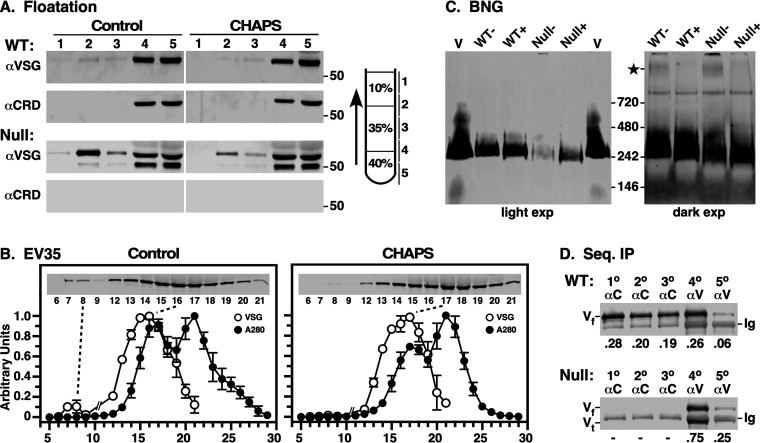
FIG 3.
Characterization of shed VSG. Conditioned culture supernatants (CMs) were generated by incubating freshly harvested late-log-phase cells in fresh HMI-9 medium (6 h, 107/ml). (A) Wild-type (WT) and GPI-PLC−/− (null) conditioned culture supernatants (106 cell equivalents) were fractionated by density floatation (diagram) in the absence and presence of 16 mM CHAPS (2× CMC). Gradient fractions were analyzed by immunoblotting with anti-VSG and anti-CRD antibodies. Vertical white lines indicate sections that were digitally excised for presentation after image processing. Mobility of molecular mass markers is indicated (right). WT versus null data are from different gels/images, and can only be compared qualitatively. (B) Gel filtration of WT conditioned medium. CM (0.5 ml) was gravity applied to an EV35 column, and 0.5-ml fractions were collected from T0. The A280 of each fraction was determined, and VSG was assayed by immunoblotting (inset). Note that fractions 10 and 11 were omitted. Each run was normalized to the fraction with the highest value, and data are presented as means ± SDs (n = 3). For CHAPS treatment, load samples were adjusted to 16 mM CHAPS and run in buffer with 1 mM CHAPS. (C) CM plus (+) or minus (−) 1% dodecylmaltoside (DDM) was fractionated by blue native gel electrophoresis and analyzed by immunoblotting. Purified sVSG (V; MITat1.2) was run as a control. A darker exposure of the CM lanes is presented on the right. DDM-sensitive high-molecular-weight VSG is indicated (star). Note that native VSG consistently migrates at approximately twice its known size (∼110 kDa) relative to the manufacturer supplied markers. This effect, which is likely due to the highly elongated and glycosylated nature of VSGs in general, has been noted by others (46). (D) WT and null CMs were subjected to sequential immunoprecipitation, 3× with anti-CRD (1°, 2°, and 3°) and then twice with anti-VSG (4° and 5°) antibodies. Precipitates were analyzed by immunoblotting with anti-VSG antibody. Mobilities of full-length (Vf) and truncated (Vt) VSG and background immunoglobulin heavy chain (Ig) are indicated. Fractional recoveries for each precipitation are shown (bottom).

