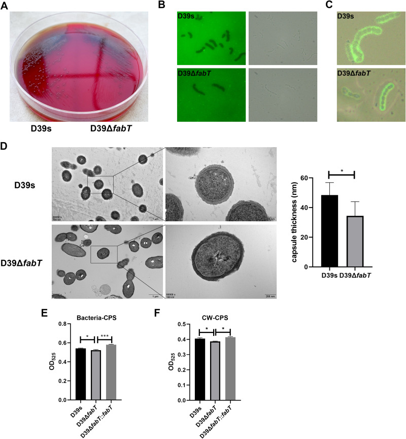
FIG 1.
Deletion of fabT leads to decreased capsule production. (A) Colony morphology on a blood agar plate (BAP) of D39s (left) and D39ΔfabT (right) strains. (B) Fluorescence and bright-field microscopy of D39s (upper panel) and D39ΔfabT (lower panel) in the presence of FITC-dextran (×100 objective). (C) Overlay of bright-field and fluorescence microscopy of D39s and D39ΔfabT mutant strains showing the capsule in green (anti-type 2 capsule antibodies and FITC-goat anti-rabbit IgG). (D) Transmission electron microscopy of D39s and D39ΔfabT. The mean capsule layer diameters are indicated; n = 20. (E and F) Comparisons of whole-cell and cell wall-associated CPS using the uronic acid assay. Statistical analysis was performed using the unpaired t test. *, P < 0.05; **, P < 0.01; ***, P < 0.001.

