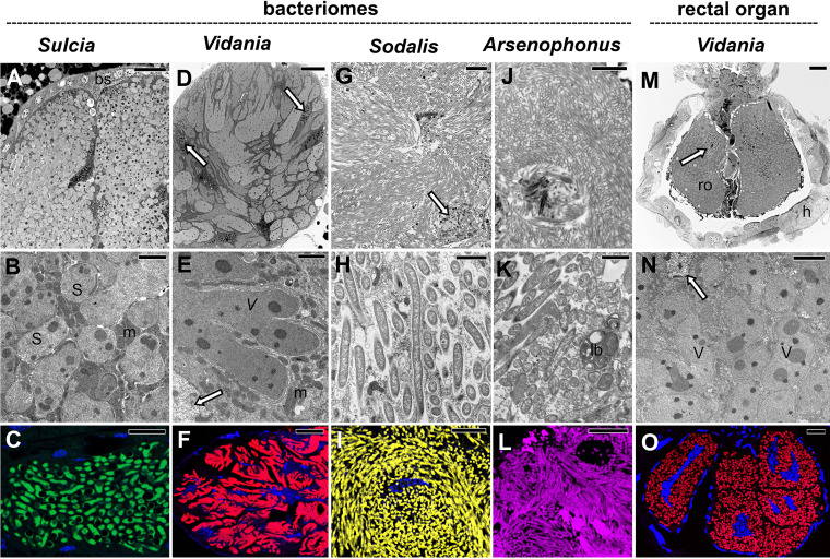
FIG 4.
Tissue localization and morphology of symbionts in the Dictyopharidae species examined. Top row (A, D, G, J, and M): the organization of symbionts within bacteriomes or the rectal organ. Light microscopy (LM), bar = 20 μm. Middle row (B, E, H, K, and N): the ultrastructure of symbiont cells. Transmission electron microscopy (TEM), bar = 2 μm. Bottom row (C, F, I, L, and O): fluorescence in situ hybridization (FISH) microphotographs of symbiont cells within the bacteriome or rectal organ. Probes specific to each of the symbionts were used. Blue represents cell nuclei stained with DAPI. Confocal laser microscope (CLM), bar = 20 μm. Insect species: (A) D. multireticulata, (B and L) R. scytha, (C, E to I, M, and N) C. krueperi, (D and O) D. pannonica, (J and K) D. europaea. Abbreviations and symbol: bs, bacteriome sheath; h, hindgut; lb, lamellar body; m, mitochondrion; ro, rectal organ; S, Sulcia; V, Vidania; white arrow, bacteriocyte nucleus.

