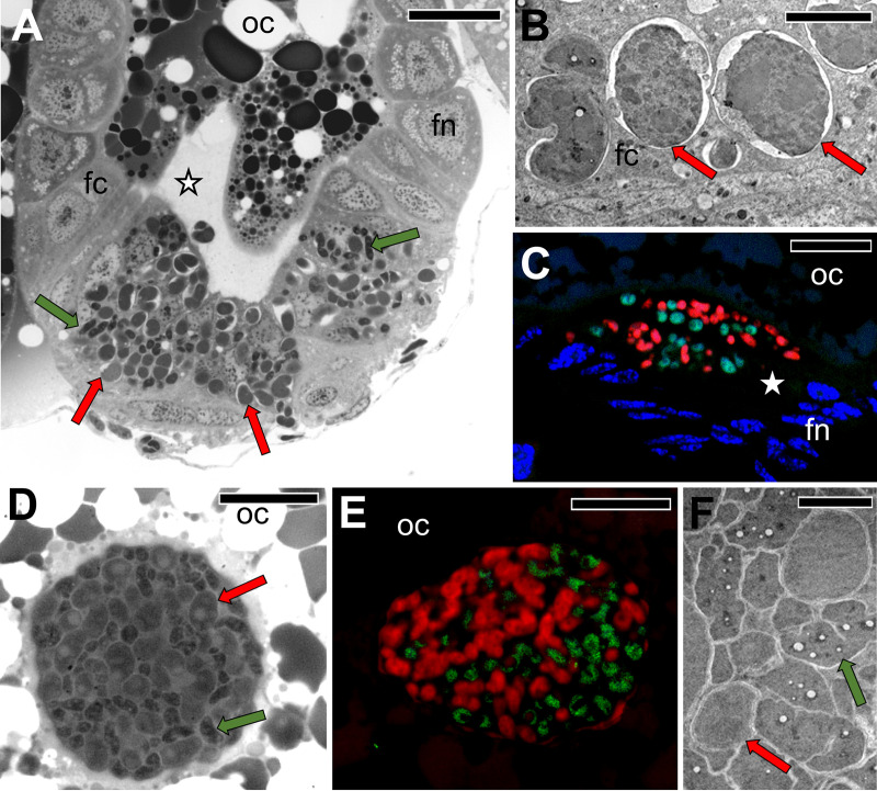
FIG 8.
Transovarial transmission of Sulcia and Vidania into the posterior end of the ovariole in representatives of Dictyopharinae subfamily. (A) The migration of Sulcia and Vidania to the perivitelline space through the follicular epithelium surrounding the posterior pole of the terminal oocyte. D. multireticulata, LM, bar = 20 μm. (B) Vidania in the cytoplasm of the follicular cell. D. pannonica, TEM, bar = 2 μm. (C) Accumulation of Sulcia (green) and Vidania (red) in the perivitelline space. C. krueperi. Confocal microscope, bar = 20 μm. (D) A “symbiont ball” containing Sulcia and Vidania in the deep depression of the oolemma at the posterior pole of the terminal oocyte. D. multireticulata, LM, bar = 20 μm. (E) In situ identification of symbionts in the “symbiont ball” in the mature oocyte of C. krueperi. CLM, bar = 20 μm. (F) Fragment of “symbiont ball.” D. multireticulata, TEM, bar = 2 μm. Abbreviations and symbols: fc, follicular cell; fn, the nucleus of a follicular cell; oc, oocyte; asterisk, perivitelline space; red cells/arrows, Vidania; green cells/arrows, Sulcia.

