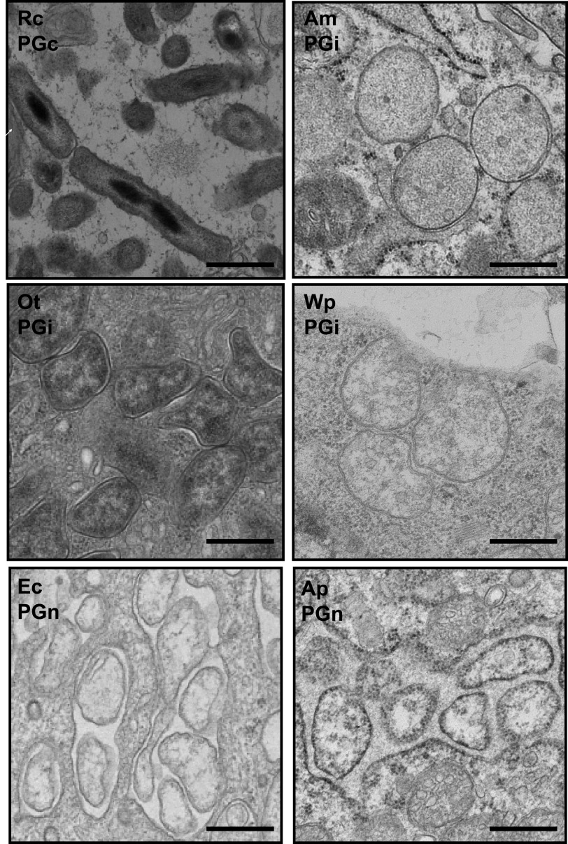
FIG 3.
Transmission electron microscopy analysis of PGc, PGi, and PGn Rickettsiales. Bacteria were grown in host cells and prepared for fixed thin-section transmission electron microscopy. Micrographs show that the PGi R. canadensis is rod-shaped compared with PGi and PGn Rickettsiales. PGi and PGn bacteria are round and/or pleiomorphic, reflecting the low level or absence of PG in their cell walls.

