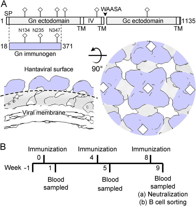
FIG 1.
HTNV Gn immunization strategy. (A) (Upper) Schematic diagram illustrating the Gn and Gc glycoproteins encoded in the HTNV M segment. The construct of HTNV Gn (residues 18 to 371) used for immunization is highlighted and colored lilac (produced with DOG 4.0 [94]). Predicted N-linked glycosylation sites (NXT/S, where X≠P) are annotated with sticks. (Lower) Schematic diagram of the (Gn-Gc)4 lattice (based upon EMD-4867), as revealed by previous cryo-ET and X-ray crystallography studies (35). Although the Gc may likely impinge, the N-terminal region of the hantaviral Gn is predicted to make up the majority of the membrane-distal region (lilac) of the (Gn-Gc)4 lattice. (B) Timeline of rabbit immunization experiments. Rabbits were immunized with recombinant HTNV Gn and boosted at 4-week intervals. Seven days following the third immunization, HTNV Gn binding and neutralization titers were measured. mAbs were isolated through antigen-specific single B cell sorting of PBMCs (Fig. S2).

