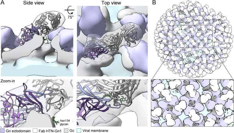
FIG 4.
Cryo-ET of HTNV VLPs in complex with Fab HTN-Gn1 provides a model for mAb-mediated obstruction of the (Gn-Gc)4 lattice. (A) Side (left) and top (right) views of the HTNV VLP−Fab HTN-Gn1 reconstruction with the crystal structure of HTNV Gn−Fab HTN-Gn1 (cartoon representation and colored as described in the legend to Fig. 3) fit into the density as a single rigid body. The HTNV VLP is shown as a surface with density corresponding to Fab HTN-Gn1 colored white, the N-terminal ectodomain of HTNV Gn colored purple, the viral membrane colored light blue, and the expected ectodomain regions of the HTNV Gc colored gray. (B) Model of Fab HTN-Gn1 binding in the context of a HTNV VLP, prepared by mapping (Gn-Gc)4 spike complexes onto the refined coordinates of a single VLP in the data set. For each position, one of the two possible overlapping binding sites was chosen randomly. Colored as described for panel A.

