
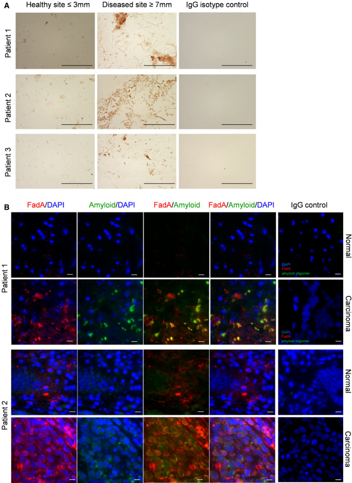
Figure 3. Amyloid‐like FadA is detected in periodontal disease and CRC.
-
ADetection of FadA in subgingival plaque samples by IHC. Three patients with periodontitis are shown here who provided plaque samples from their healthy sites (probing depth ≤ 3 mm, left panels) and periodontal diseased sites (probing depth ≥ 7 mm, middle panels). IHC was performed as described using mAb 7H7 at 1:800 dilution. Anti‐mouse IgG isotype control of the diseased site is shown (right panels). The images were taken using a 100X objective. Scale bar equals 50 μm.
-
BDouble immunofluorescent staining of paired normal and carcinoma tissues from two CRC patients. The frozen tissue sections were incubated with mAb 7H7 at 1:50 dilution and anti‐amyloid antibody A11 at 1:25 dilution, or mouse and rabbit IgG control, followed by incubation with Alexa Fluor 555‐conjugated goat anti‐mouse and Alexa Fluor 680‐conjugated donkey anti‐rabbit antibodies, both at 1:1,000 dilution. Co‐staining of FadA (red) and amyloid oligomers (green) were observed in the carcinoma, but not normal, tissues. The images were taken using a 60X objective. Scale bar equals 10 μm.