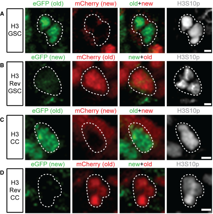
Figure EV1. Old versus new histone H3 distribution patterns in Drosophila female germline stem cells (GSCs) and cystocytes (CCs) during the first mitosis after heat shock‐induced genetic switch.

- Old (green, eGFP) versus new (red, mCherry) histone H3 patterns in a prometaphase female GSC marked by anti‐H3S10p (gray).
- Old (red, mCherry) versus new (green, eGFP) histone H3 patterns for H3Rev in a prophase female GSC marked by anti‐H3S10p (gray).
- Old (green, eGFP) versus new (red, mCherry) histone H3 patterns in a prophase female CC marked by anti‐H3S10p (gray).
- Old (red, mCherry) versus new (green, eGFP) histone H3 patterns for H3Rev in a prometaphase female CC marked by anti‐H3S10p (gray). During the first mitosis after heat shock, the new histone has not yet been globally incorporated into the chromatin, shown as low to undetectable fluorescent signals.
Data information: Scale bars: 2 µm.
