
Figure 1.
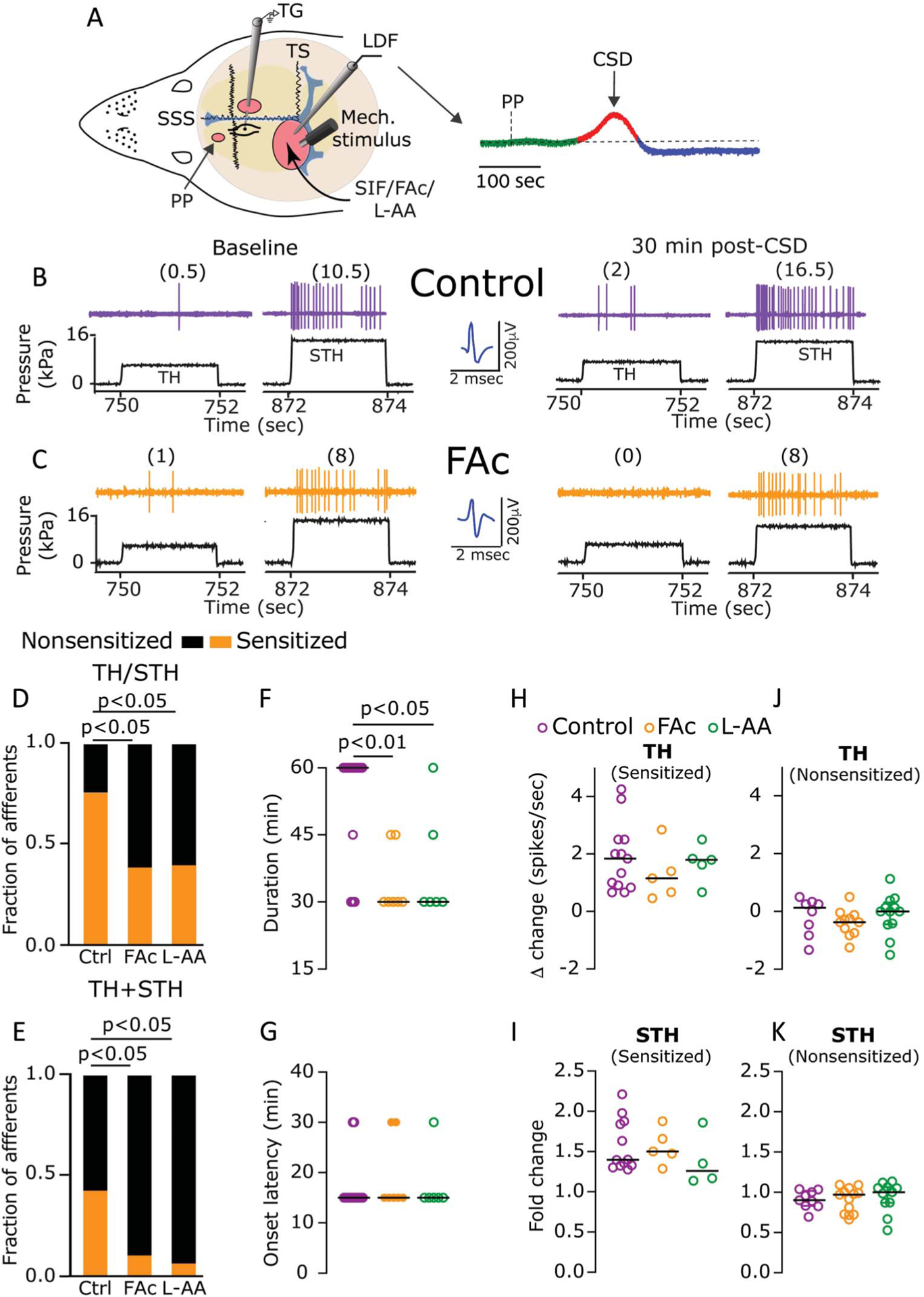
Astrocytes contribute to CSD-evoked mechanical sensitization of meningeal dural afferents. (A) Experimental setup: 3 skull openings (red ovals) were made. A small burr hole was drilled over the left frontal cortex to elicit a single CSD event using a pinprick (PP) stimulation. Afferent activity was recorded in the left trigeminal ganglion (TG) using a tungsten microelectrode inserted through a craniotomy made over the contralateral hemisphere. An ipsilateral craniotomy was made to expose a part of the left transverse sinus (TS) and superior sagittal sinus (SSS) and their vicinity to find and stimulate mechanosensitive meningeal afferents. Quantitative mechanical stimuli were delivered to the receptive field of afferents using a feedback-controlled mechanical stimulator. A laser Doppler flowmeter (LDF) probe was placed over the cortex near the receptive field of afferents to record changes in cerebral blood flow and validate CSD induction noninvasively. Induction of CSD was considered successful when the typical hemodynamic signature characterized by a large transient (~1–2 minutes) cortical cerebral hyperemia (red trace), followed by persistent (>1 hours) post-CSD oligemia (blue trace) was observed. (B and C) Examples of experimental trials depicting the responses of 2 afferents to threshold (TH) and suprathreshold (STH) mechanical stimuli (black traces) applied to their receptive field during baseline recording and then at 30 minutes after CSD elicitation in control (B) and FAc-treated animals (C). Responses in spikes/second are in parentheses. Note the sensitization in control but not after FAc treatment. When compared with controls (n = 21), the fraction of afferents that became sensitized at the TH or STH level (TH/STH, D), or at both levels (TH + STH, E) was lower in animals treated with FAc (n = 18), or L-AA (n = 15) (P < 0.05, χ2 test, treatment vs control). Inhibition of astrocytic function with FAc or L-AA also decreased the duration of the sensitization response (F); (P values indicate the Dunn post hoc test after a significant [P < 0.01] Kruskal–Wallis test between all treatments). The onset latency (G) or magnitude (H and I) of the sensitization response was not different between the 3 treatments (P > 0.05, Kruskal–Wallis). Inhibition of astrocytic function with FAc or L-AA did not affect TH or STH mechanical responses in nonsensitized afferents (J and K), (P > 0.05, Kruskal–Wallis). Data in (F–K) include all data points. Lines represent the median. CBF, cerebral blood flow; CSD, cortical spreading depression; FAc, fluoroacetate; L-AA, L-α-aminoadipate.