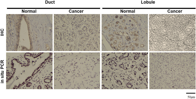
FIGURE 3.
Morphological examination of JCV T antigen in breast cancers. According to immunohistochemistry (IHC), T antigen was strongly expressed in ductal and lobular epithelial cells (brown) but positively or weakly expressed in breast ductal and lobular cancer cells. NBT/BCIP coloring is displayed as black to show positive signals in breast ductal and lobular epithelial cells by in situ PCR, while methyl green is denoted as a green color for counterstaining.

