Fig. 7.
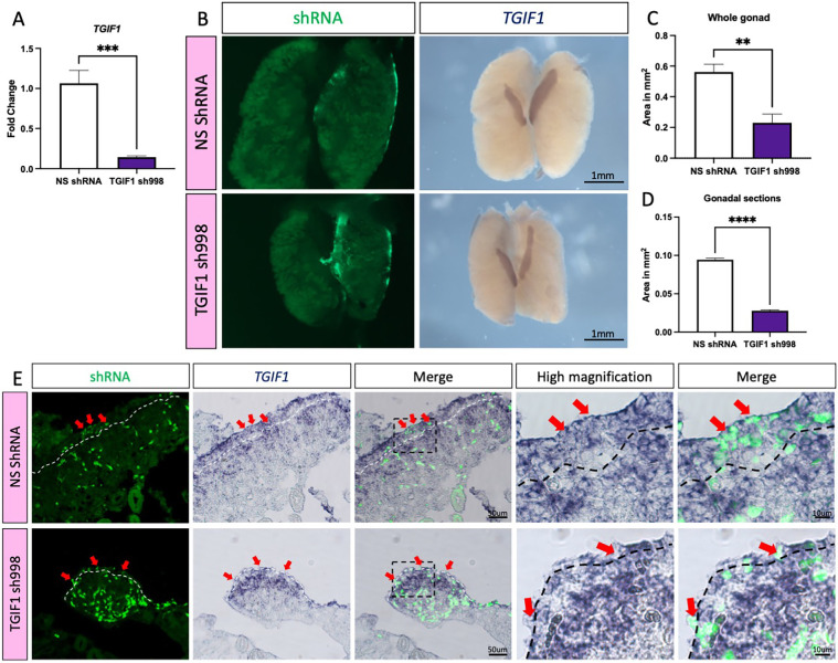
TGIF1 knockdown results in smaller ovaries. (A) DF1 cells expressing TGIF1 sh998 or non-silencing control (NS shRNA) were challenged for 48 h with RCAS(A)-GFP-T2A-TGIF1 overexpression plasmid. TGIF1 mRNA was quantified by qRT-PCR. Expression level is relative to β-actin and normalized to NS shRNA (n=6). (B) TOL2 TGIF1 knockdown (TGIF1 sh998) or non-silencing control (NS shRNA) plasmids were co-electroporated with a GFP-expressing plasmid (reporter) in female left E2.5 coelomic epithelium. Gonads were examined at E8.5 for GFP expression and TGIF1 whole-mount in situ hybridization was performed. (C) Quantification of the gonadal area (in mm2) from whole-mount images (n=5). (D) Quantification of the gonadal area (in mm2) from whole-mount gonadal sections (n=5). Bars represent mean±s.e.m. **adjusted P<0.01, ***adjusted P<0.001, ****adjusted P<0.0001. Unpaired two-tailed t-test. (E) Whole-mount sections (10 μm) were processed for immunofluorescence against GFP. Dashed white line delineates the gonadal epithelium. Red arrows indicate GFP-positive (shRNA-targeted) epithelial/cortical cells.

