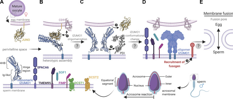
Figure 3.
Current model of sperm–egg attachment and fusion. (A) Acrosome reaction. After the acrosome reaction, IZUMO1 (blue), SPACA6 (purple), and TMEM95 (violet) colocalize to the equatorial regions of sperm. FIMP (pink) appears to function before the acrosome reaction. There are conflicting data on whether or not TMEM95 interacts with IZUMO1. SOF1 (turquoise) is a secreted sperm protein. DCST1 (green) and DCST2 (orange) are transmembrane proteins implicated in regulating the protein stability of SPACA6. (B) Initial attachment. After the sperm reaches the PVS, it attaches to the egg. IZUMO1 is localized on the equatorial segment of acrosome-reacted sperm and its counterpart receptor, JUNO (yellow), on the oocyte membrane. JUNO specifically recognizes and binds to IZUMO1 in a monomeric conformation. IZUMO1 binding to JUNO drives the accumulation of CD9 (pink) at the sperm–egg interface to form a physical anchor that holds the sperm and oocyte membranes in proximity. (C) IZUMO1 multimerization. After the initial IZUMO1–JUNO attachment, the complex undergoes a dimerization event. The trigger for IZUMO1 oligomerization is not fully understood; however, colocalization analysis revealed the presence of PDI (gray) on the sperm surface. JUNO is thought to be shed from the oolemma and into the PVS after fertilization. (D) Fusogen recruitment. The bona fide sperm–egg fusogen remains a mystery. However, data suggest that IZUMO1 forms a scaffold to recruit the gamete fusion complex. The roles of SPACA6, TMEM95, and SOF1 remain unclear, but these proteins likely play roles in fusion. (E) Fusion pore formation. The merger of the egg and sperm membranes requires modulation of the membrane architecture. The fusogen is thought to catalyze the formation of a hemifusion intermediate, which is a stalk-like structure where the outer leaflets of the sperm and egg membrane bilayers mix. Subsequently, the inner bilayer leaflets mix to form the fusion pore. The precise mechanism of this step will require the identification of the sperm–egg fusogen. Created with BioRender.

