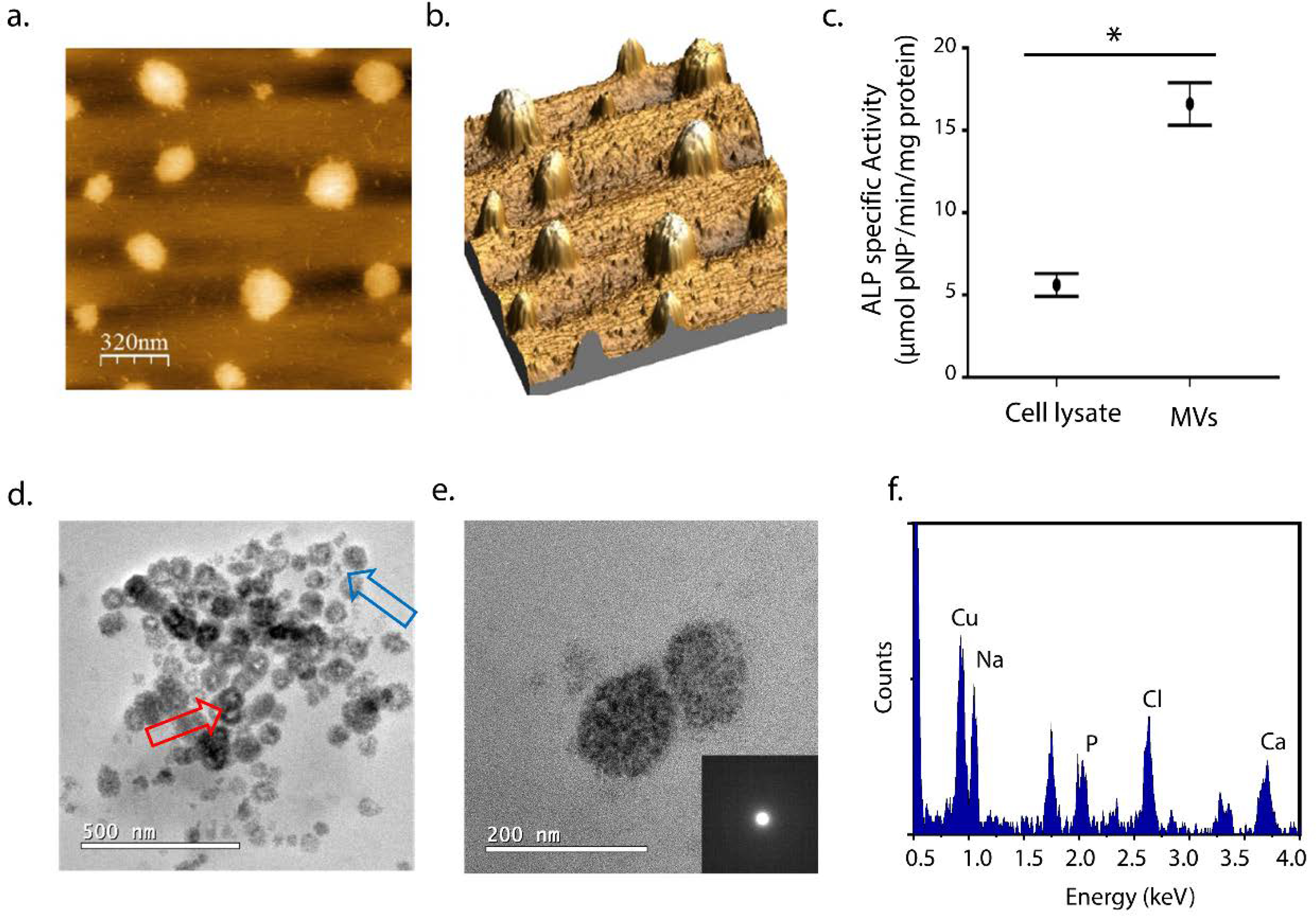
Figure 1.

Characterization of MVs extracted from chicken embryo femurs: (a) AFM images by height distribution and (b) 3D projection of the isolated MVs on mica. (c) ALP specific activity for cell lysate and MVs fractions (μmol pNP−/min/mg protein). Values are presented as mean ± standard error and statistical significance was assessed with t-tests (*p < 0.05). (d) TEM images of the MVs after 24 h of mineralization in SCL (2 mM CaCl2 and 3.42 mM NaH2PO4) at 37°C. (e) Enlarged TEM image of a pair of mineralized-vesicles and their respective amorphous-like SAED and (f) the respective EDS spectrum indicating the presence of Ca and P. The presence of Na and Cl is also observed due to the large amount of NaCl in the SCL and Cu is from de TEM grid.
